IMAGE

Fig. 5

ID
ZDB-IMAGE-200907-11
Source
Figures for Zhou et al., 2019
Image
Figure Caption

Fig. 5

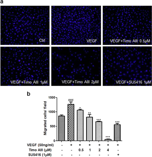
Timo AIII inhibits VEGF-induced migration in HUVECs. The migration ability of HUVECs was measured by a classical transwell migration assay. a HUVECs were treated with VEGF (50 ng/mL) and various concentrations (0.5, 1, 2, and 4 µM) of Timo AIII for 24 h. HUVECs incubated with 0.1% DMSO served as a vehicle control and with SU5416 (1 µM) as a positive control. The nuclei of HUVECs were stained with Hoechst 33342 and produced blue fluorescence when excited by the UV light. Representative images show the migration ability of HUVECs under the different treatment conditions. b The statistical graph shows the quantitative analysis of HUVEC migration. Data are presented as the cell number per field. The results are presented as the means ± S.E.M. ###P < 0.001 versus the control group; *P < 0.05, **P < 0.01, and ***P < 0.001 versus the VEGF-treated group

Acknowledgments
This image is the copyrighted work of the attributed author or publisher, and ZFIN has permission only to display this image to its users. Additional permissions should be obtained from the applicable author or publisher of the image. Full text @ Acta Pharmacol. Sin.