
Figure 2
- ID
- ZDB-IMAGE-200903-2
- Publication
- Tsai et al., 2020 - Newly identified Gon4l/Udu-interacting proteins implicate novel functions
- All Figures
- Figures for Tsai et al., 2020
|
Figure 2
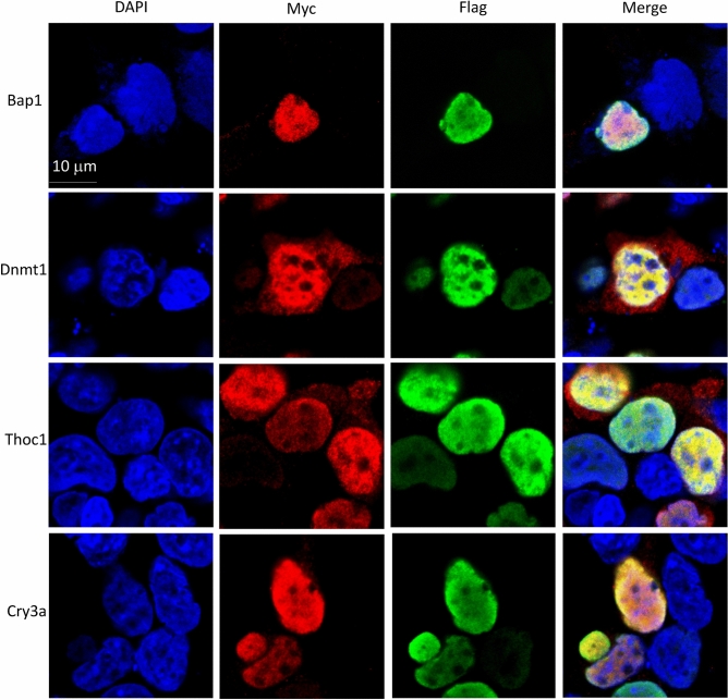
Confocal images for the cellular co-localization analysis of Udu and its interacting proteins. For co-localization analysis, Flag-tagged Udu-P/S construct was co-transfected individually with each Myc-tagged construct, including Bap1, Dnmt1, Thoc1, and Cry3a. Anti-FLAG M2 monoclonal antibody and rabbit anti-MYC antibody were used. Alexa Fluor 488-conjugated anti-mouse secondary antibody and Alexa Fluor 564-conjugated anti-rabbit secondary antibody were then applied. Green fluorescence indicates Udu-P/S, while red fluorescence indicates the expression of Myc-tagged proteins. Blue color is DAPI used for nuclear counterstain. Merged images show the nuclear co-localization of Udu and the interacting proteins.