IMAGE

Figure 6.

ID
ZDB-IMAGE-200829-70
Antibodies
Source
Figures for Liu et al., 2020
Image
Figure Caption

Figure 6.

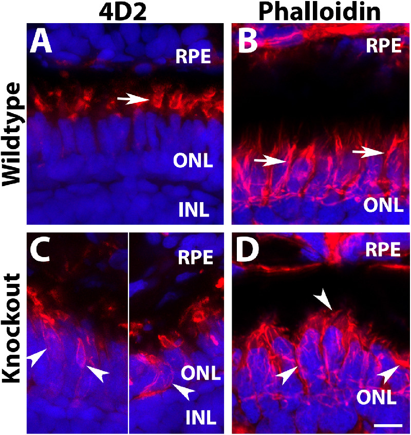
Defective organization of F-actin in tmem216 knockout photoreceptors. Cryosections were immunostained with 4D2 (red). Some sections were stained with Phalloidin-RITC for F-actin (red). (A, C) 4D2 staining of wildtype and tmem216snyΔ175 homozygous retina. 4D2 labels rod outer segments in the wildtype zebrafish (arrow in A); however, its reactivity was frequently mislocalized to the rod cell body in the knockouts (arrowheads in C). (B, D) Phalloidin staining in wildtype and tmem216snyΔ175 homozygous retina. Vertically oriented F-actin in the wildtype (arrows in B) were disrupted in tmem216snyΔ175 homozygous retina (arrowheads in D). Scale bar in H: 6 µm.

Figure Data
Acknowledgments
This image is the copyrighted work of the attributed author or publisher, and ZFIN has permission only to display this image to its users. Additional permissions should be obtained from the applicable author or publisher of the image. Full text @ Invest. Ophthalmol. Vis. Sci.