
|
Figure 1
Recording from RGC Dendrites and Somata
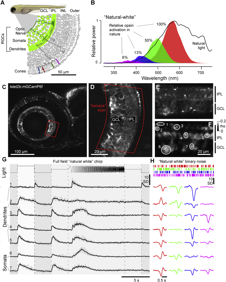
(A) Schematic of Islet2b:mGCaMP6f expression in RGCs (green) across a section of the larval zebrafish eye, with somata in the ganglion cell layer (GCL) and dendrites in the inner plexiform layer (IPL); see also
(B) Average spectrum of natural daylight measured in the zebrafish natural habitat from the fish’s point of view along the underwater horizon (solid line). Convolution of the zebrafish’s four cone action spectra with this average spectrum (shadings) was used to estimate the relative power each cone surveys in nature, normalized to red cones (100%). Stimulation LED powers were relatively adjusted accordingly (“natural white”).
(C and D) GCaMP6f expression under two-photon surveyed across the entire eye’s sagittal plane (C) and zoom-in to the strike zone as indicated (D). Within the zoomed field of view, a curved scan path was defined (“banana scan”) to follow the curved GCL and IPL for activity recordings (E), which effectively “straightened” the natural curvature of the eye.
(E and F) Example activity scan with RGC dendrites occupying the top part of the scan in the IPL and somata occupying the bottom part in the GCL as indicated (E) and correlation projection [
(G) Mean (black) and individual repeats (gray) example responses of ROIs from (E) to full-field stimulation as indicated.
(H) As (G), now showing linear kernels to red, green, blue, and UV components recovered from natural white noise stimulation (
Note that several ROIs display a robust UV component despite the ~20-fold attenuated stimulation power in this band relative to red (B). See also