Figure Caption
Fig 8
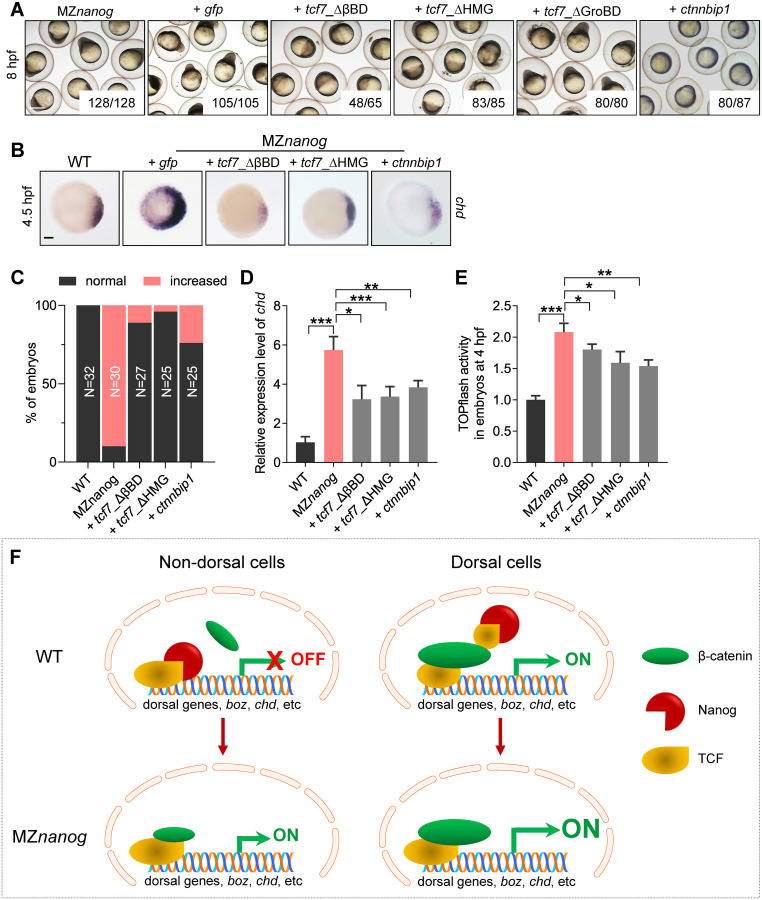
Confrontation of the β-catenin transcriptional activity in nucleus rescues the developmental defect of MZ<italic>nanog</italic>.(A) Injection of tcf7)_ΔβBD, tcf7_ΔHMG, or ctnnbip1 mRNA rescued the early developmental defects of MZnanog, whereas overexpression of tcf7_ΔGroBD did not. Phenotypes were observed at 8 hpf. At least 50 embryos were injected, and 3 independent experiments were performed. The numbers below the morphology pictures mean number of embryos showing representative phenotype/total number of embryos. Scale bar, 500 μm. (B) WISH analysis showing excessive and ectopic expression of chd in MZnanog was rescued by overexpression of tcf7)_ΔβBD, tcf7_ΔHMG, or ctnnbip1 at 4.5 hpf. Scale bar, 100 μm. (C) Statistical analysis of the embryos in panel B. N represents analyzed embryo number. (D) Relative mRNA level of chd in embryos of WT, MZnanog, and MZnanog injected with tcf7)_ΔβBD, tcf7_ΔHMG, or ctnnbip1 mRNA at 4.5 hpf examined by RT-qPCR. Error bars, mean ± SD, *P < 0.05, **P < 0.01, ***P < 0.001. (E) Relative β-catenin transcriptional activity in embryos of WT, MZnanog, and MZnanog injected with tcf7)_ΔβBD, tcf7_ΔHMG, or ctnnbip1 mRNA at 4 hpf examined by TOPflash assay. Error bars, mean ± SD, *P < 0.05, **P < 0.01, ***P < 0.001. (F) The model of Nanog repressing β-catenin transcriptional activity in nondorsal cell nuclei in WT embryo, and the ectopic activation of β-catenin transcriptional activity in the absence of Nanog in MZnanog embryo. In nondorsal cells of WT embryo, the amount of nucleus-deposited maternal Nanog (red cartoon object) is much higher than that of the nuclear β-catenin (green cartoon object); therefore Nanog binds to TCF (yellow cartoon object), and the β-catenin transcriptional activity is not activated. In nondorsal cells of MZnanog embryo, however, because of the absence of nanog in the nuclei, the small amount of nuclear β-catenin binds to TCF to activate the expression of dorsal genes (boz, chd, etc.), resulting in hyperdorsalization of the embryo. In dorsal cells of WT embryo, the amount of nuclear β-catenin is much higher than Nanog and facilitates the formation of β-catenin-TCF transcriptional complex to induce the expression of dorsal genes, boz, chd, etc. The P values in this figure were calculated by Student t test. The underlying data in this figure can be found in S1 Data. hpf, hours post fertilization; MZnanog, maternal zygotic mutant of nanog; RT-qPCR, reverse-transcription quantitative PCR; tcf7)_ΔβBD, β-catenin-binding domain deleted Tcf7, tcf7_ΔHMG, high mobility group (LEF1-binding domain) deleted Tcf7; tcf7_ΔGroBD, Groucho-binding domain deleted Tcf7; WISH, whole-mount in situ hybridization; WT, wild type.
Acknowledgments
This image is the copyrighted work of the attributed author or publisher, and
ZFIN has permission only to display this image to its users.
Additional permissions should be obtained from the applicable author or publisher of the image.
Full text @ PLoS Biol.