
Figure 3
- ID
- ZDB-IMAGE-200725-4
- Publication
- Poplimont et al., 2020 - Neutrophil Swarming in Damaged Tissue Is Orchestrated by Connexins and Cooperative Calcium Alarm Signals
- All Figures
- Figures for Poplimont et al., 2020
|
Figure 3
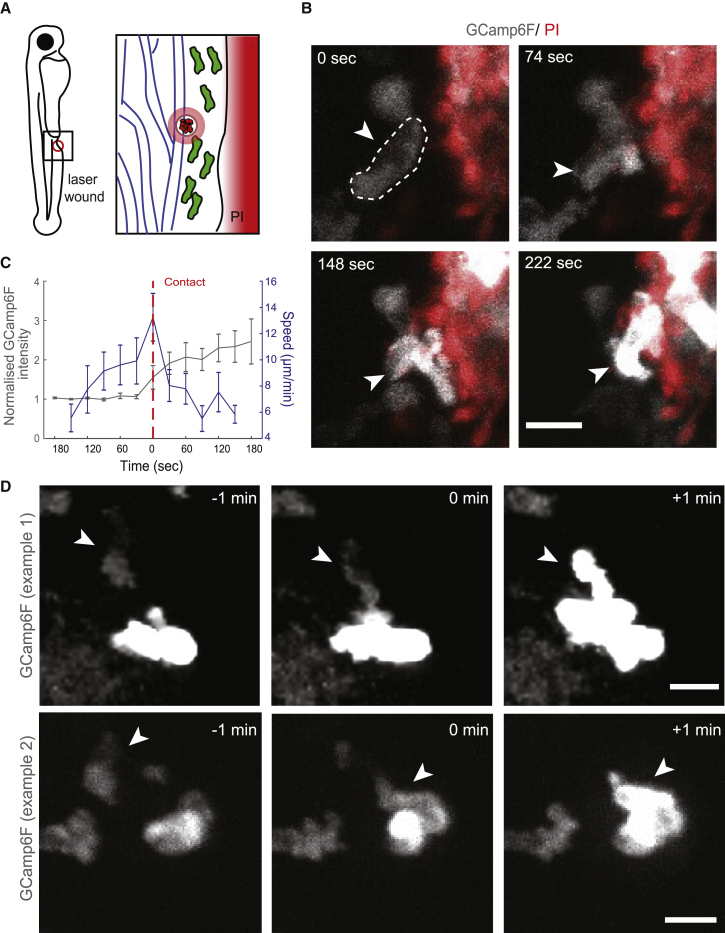
5-LO-Associated Calcium Fluxes Are Triggered upon Contact with Necrotic Cells or Neutrophils with Ongoing Fluxes
(A) Schematic of two-photon laser wounding in the presence of propidium iodide (PI).
(B) Time-lapse, two-photon confocal projection images of a GCamp6F-expressing (white) neutrophil (indicated with an arrow) entering a contact with PI+ cells/tissue (red) in a Tg(
(C) Quantification of speed (blue) and normalized GCamp6F (gray) in neutrophils before and after contact with PI+ tissue. GCamp6F intensity was normalized as in
(D) Examples of cell contacts transmitting calcium fluxes. Each case is represented by time-lapse images of a non-calcium-fluxing neutrophil (arrow) contacting a fluxing neutrophil. Time in minutes is indicated relative to cell-cell contact. Scale bar represents 10 μm. The quantification of neutrophil transmission of calcium fluxes is indicated in
Error bars represent SEM. See also