IMAGE

Figure 2

ID
ZDB-IMAGE-200716-80
Source
Figures for Chen et al., 2020
Image
Figure Caption

Figure 2

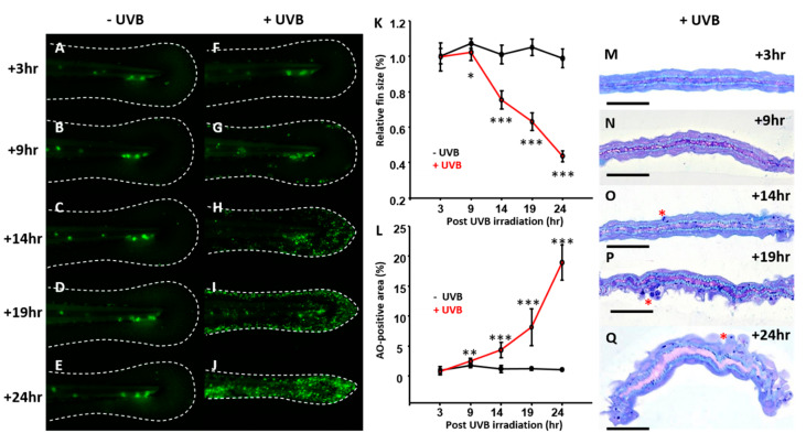
Time course changes of caudal fin in zebrafish after UVB irradiation. Zebrafish embryos aged 3 dpf were irradiated with UVB at doses of 300 J/m2. (FJ) The embryos were then collected at 3, 9, 14, 19, and 24 h after irradiation and subjected to AO staining to detect apoptosis and measuring the relative size of caudal fin. The control group was for comparison (AE). (K) Dynamic changes of the relative caudal fin size in zebrafish with UVB irradiation at dose of 300 J/m2. (L) Dynamic changes of the relative apoptotic cells with UVB irradiation at a dose of 300 J/m2. (MQ) Semi-thin sectioning was conducted to compare the interior morphology of caudal fin under UVB irradiation at a dose of 300 J/m2. (n = 5–15. Student’s t-test was performed on data from experiments. * p < 0.05, ** p < 0.01, *** p < 0.001). Scale bar = 25 μm in (MQ).

Acknowledgments
This image is the copyrighted work of the attributed author or publisher, and ZFIN has permission only to display this image to its users. Additional permissions should be obtained from the applicable author or publisher of the image. Full text @ Animals (Basel)