IMAGE

Figure 1

ID
ZDB-IMAGE-200716-79
Source
Figures for Chen et al., 2020
Image
Figure Caption

Figure 1

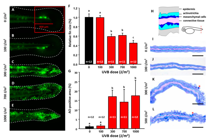
Optimization of UVB irradiation dosage to induce caudal fin damage in zebrafish. Zebrafish embryos aged 3 dpf were exposed to UVB irradiation at different doses of 0 (A), 100 (B), 300 (C), 700 (D), and 1000 (E) J/m2. Twenty-four hours post irradiation, embryos were stained with acridine orange (AO) to detect apoptosis and the relative size of caudal fin was calculated and compared. (F) Quantitative measurement of caudal fin area under different doses of UVB irradiation. (G) Quantitative measurement of cell apoptosis under different doses of UVB irradiation. The apoptosis was detected by AO staining. One-way ANOVA test was conducted to determine the significance. Column with the same label indicates statistic insignificance. Semi-thin section was conducted to compare the morphology in caudal fin at different doses from 0 (I), 100 (J), 300 (K), and 500 (L) J/m2. The cellular organization of a cross-section of caudal fin is illustrated in (H). Scale bar = 25 um in (IL).

Acknowledgments
This image is the copyrighted work of the attributed author or publisher, and ZFIN has permission only to display this image to its users. Additional permissions should be obtained from the applicable author or publisher of the image. Full text @ Animals (Basel)