IMAGE

FIGURE 2

ID
ZDB-IMAGE-200702-17
Source
Figures for Otten et al., 2020
Image
Figure Caption

FIGURE 2

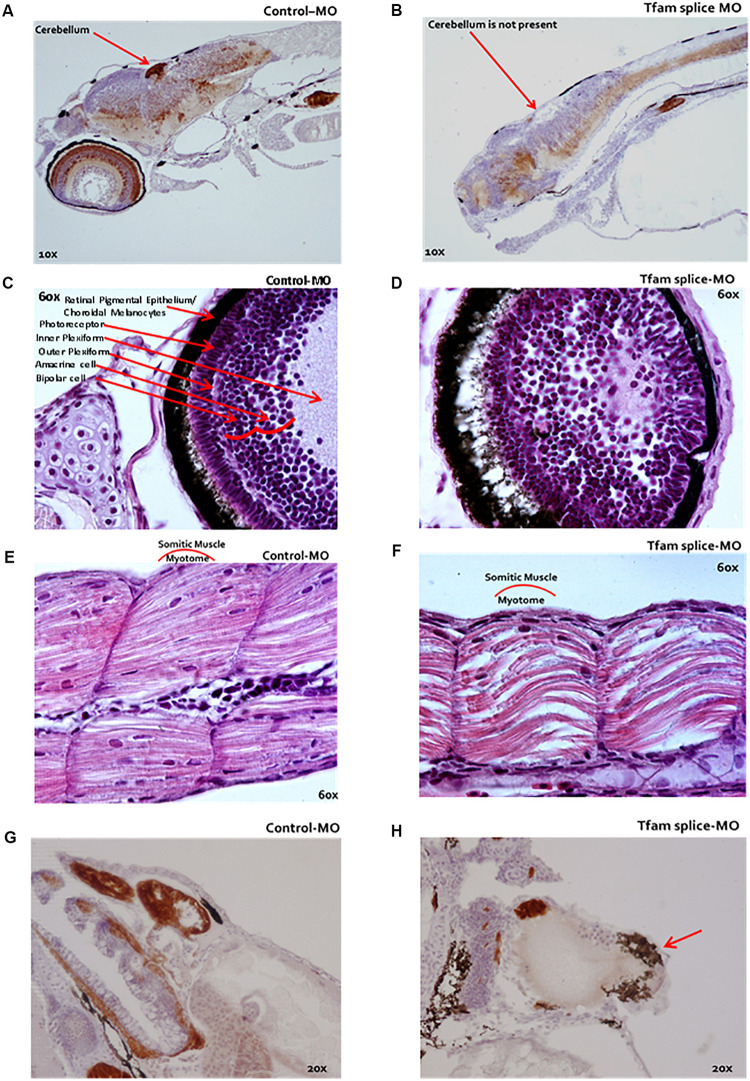
Microscopy of zebrafish embryos following TFAM knockdown. (A–H) Microscopic analysis following serial HE-staining of zebrafish embryos at 4 dpf, which were injected at 1 hpf with either 2 ng tfam splice-MO (B–E) or 2 ng Ctrl-MO, (A,C,E,G)n = 13 each condition. (B) The cerebellum was missing in the phenotype or a delayed development of this cerebellum in these fish. (D) The eyes were smaller and the eye layers were less developed. (F) A muscle phenotype was clearly visible in the dorsal fin, especially in the myotomic area, which was less compact in these somatic muscles. (H) The fish displayed a marked pericardial oedema (red arrow). They did not develop a normally looped heart as in Ctrl-MO (G), but the hearts were dilated in the phenotypes (H).

Acknowledgments
This image is the copyrighted work of the attributed author or publisher, and ZFIN has permission only to display this image to its users. Additional permissions should be obtained from the applicable author or publisher of the image. Full text @ Front Cell Dev Biol