IMAGE

FIGURE 4

ID
ZDB-IMAGE-200614-21
Source
Figures for Van Der Meulen et al., 2020
Image
Figure Caption

FIGURE 4

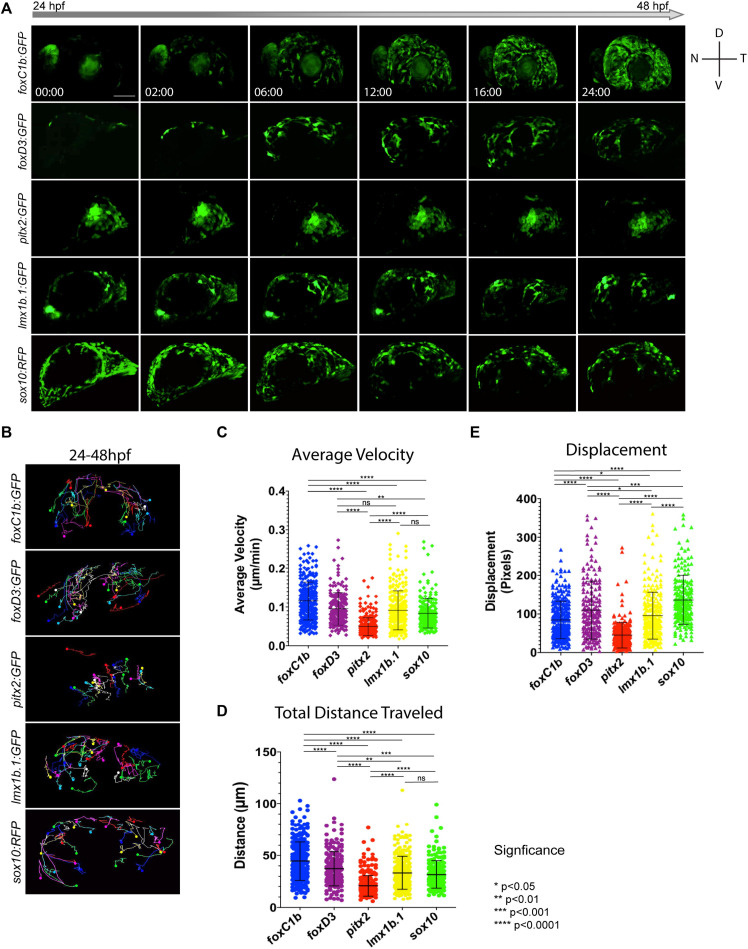
In vivo 4D imaging of POM anterior segment colonization. (A) 4D in vivo imaging of the AS conducted between 24–48 hpf using POM transgenic lines. Time stamp hours:minutes. Scale bar = 50 μm. (B) Individual cell tracking for each POM transgenic line reveals migratory patterns during early AS colonization. Solid spheres indicate terminal end of track. (C–E) Cell tracking measurements of average migratory velocity (ANOVA p < 0.0001), total migration distance (ANOVA p < 0.0001), and migratory displacement within the AS (ANOVA p < 0.0001).

Acknowledgments
This image is the copyrighted work of the attributed author or publisher, and ZFIN has permission only to display this image to its users. Additional permissions should be obtained from the applicable author or publisher of the image. Full text @ Front Cell Dev Biol