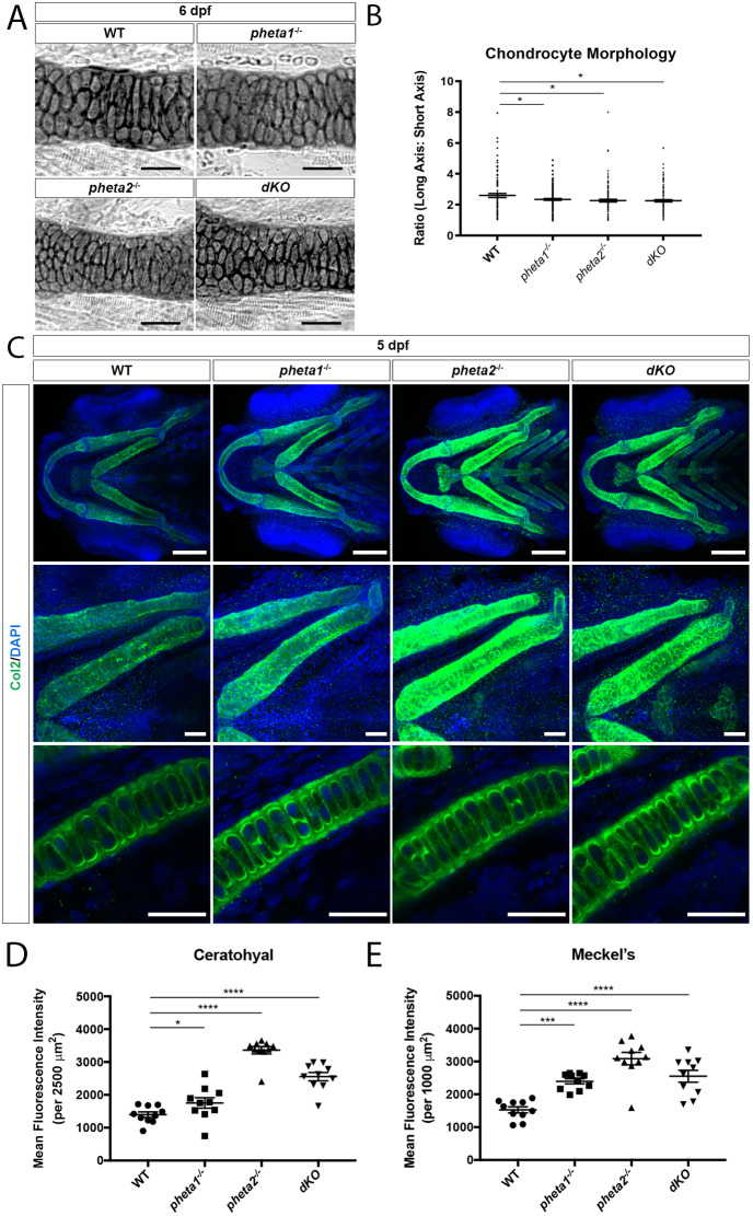
Loss of pheta1/2 disrupts chondrocyte maturation. (A) Ceratohyal cartilage, stained with Alcian Blue in flat-mount preparation. Scale bars: 50 µm. (B) Chondrocyte morphology analysis in ceratohyal cartilage (within a 200 µm2 area). (C) Top row: Z-projection ventral view of larvae immunostained for Col2 (green). Nuclei are labeled with DAPI (blue). Scale bars: 100 μm. Middle row: higher-magnification Z-projection images of corresponding ceratohyal cartilage. Scale bars: 25 μm. Bottom row: higher-magnification single optical section images of corresponding ceratohyal cartilage, depicting extracellular secretion of type II collagen. Scale bars: 25 µm. (D,E) Quantification of mean fluorescence intensity in the ceratohyal (D) and Meckel's cartilage (E) in 5 dpf larvae. *P<0.05, ***P<0.001, ****P<0.0001.
This image is the copyrighted work of the attributed author or publisher, and
ZFIN has permission only to display this image to its users.
Additional permissions should be obtained from the applicable author or publisher of the image.
Full text @ Dis. Model. Mech.
Your Input Welcome
Thank you for submitting comments. Your input has been emailed to ZFIN curators who may contact you if
additional information is required.
Oops. Something went wrong. Please try again later.