
Fig. 8
- ID
- ZDB-IMAGE-200514-14
- Genes
- Antibodies
- Publication
- Koth et al., 2020 - Runx1 promotes scar deposition and inhibits myocardial proliferation and survival during zebrafish heart regeneration
- All Figures
- Figures for Koth et al., 2020
|
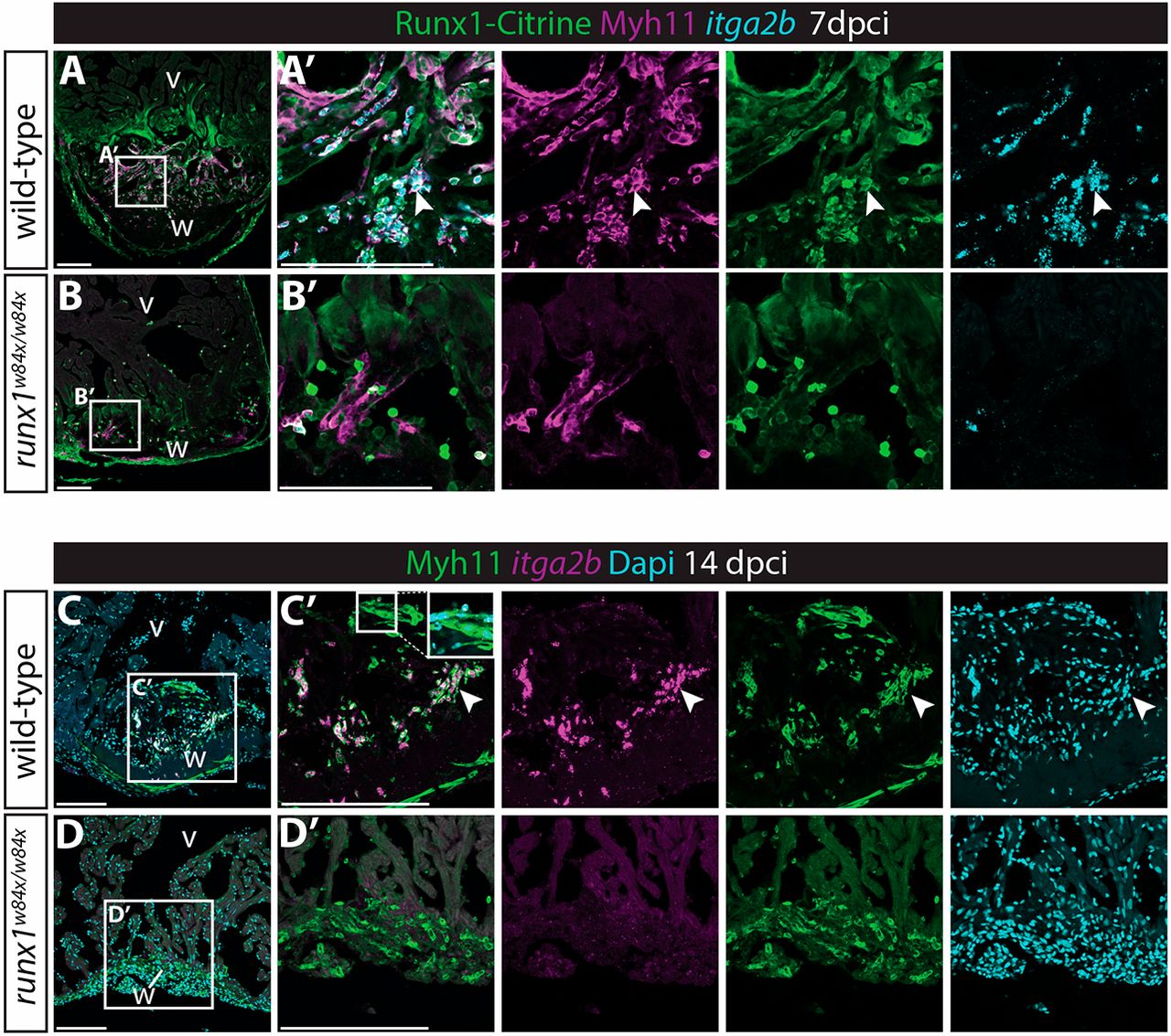
Fig. 8 Myh11-positive endocardial cells and thrombocytes retain their double identity. (A-B′) In situ hybridisation for itga2b combined with immunohistochemistry for Citrine and Myh11. Arrowheads indicate Myh11-positive itga2b-positive thrombocytes present in the wild-type wound that are largely missing in the runx1 mutant wound at 7 dpci. (C-D′) In situ hybridisation for itga2b combined with immunohistochemistry for Myh11 with nuclear Dapi staining. Both the endocardium (inset) and thrombocytes (arrowheads) still express Myh11 in the wild-type wound at 14 dpci, while being absent in the mutant wound. v, ventricle; w, wound. Scale bars: 100 μm.