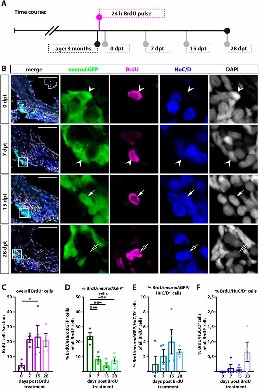
Fig. 4 neurod:GFP-positive cells are proliferating and give rise to new neurons at juvenile stages. (A) Time course of the BrdU pulse chase experiment: 3-month-old Tg(neurod:GFP) zebrafish were sacrificed at 0, 7, 15 and 28 dpt. (B) Antibody staining shows BrdU/neurod:GFP-positive cells (0 and 7 dpt, arrowheads) which differentiate into BrdU/neurod:GFP/HuC/D-positive (15 dpt, solid arrows) and BrdU/HuC/D-positive neurons (28 dpt, open arrows). Panels on right show magnification of boxed areas in left panels. (C-F) Quantification of antibody staining. (C) The overall number of BrdU-positive cells increases from 0 dpt to later time points, indicating continued proliferation of precursor cells. (D) Percentage of BrdU/neurod:GFP-positive cells of all BrdU-positive cells decreases significantly from 0 dpt to later time points. (E) At 7 dpt, some BrdU/neurod:GFP-positive cells have already differentiated into immature BrdU/neurod:GFP/HuC/D-positive cells. (F) First newborn BrdU/HuC/D-positive neurons appear as early as 7 dpt and the percentage of BrdU/HuC/D positive neurons increases over time. Scale bars: 50 µm. Cross-sections show dorsal to the top and lateral to the right. For all quantifications n=4 (n=fish; 1 SAG/fish; 12 sections/SAG); data are presented as mean±s.e.m. Ordinary one-way ANOVA with Tukey's multiple comparison test. *P≤0.05; ***P≤0.001.
Image
Figure Caption
Acknowledgments
This image is the copyrighted work of the attributed author or publisher, and
ZFIN has permission only to display this image to its users.
Additional permissions should be obtained from the applicable author or publisher of the image.
Full text @ Development

