IMAGE

Figure 3.

ID
ZDB-IMAGE-200430-8
Source
Figures for Woodard et al., 2019
Image
Figure Caption

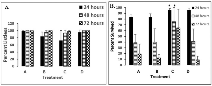
Figure 3. Shows percentages of 5 dpf demonstrating either listless (A.) or survival (B.) phenotypes under various treatment conditions after 24–72 hours of exposure. Error bars represent ± SD. Lettered groups of bars represents the following treatments: A—50 µM nNOSI; B—500 µM 6-OHDA; C—6-OHDA + nNOSI co-treatment; D—6-OHDA + DETA-NO co-treatment. Asterisk = <italic>p</italic> < 0.01 between groups.

Acknowledgments
This image is the copyrighted work of the attributed author or publisher, and ZFIN has permission only to display this image to its users. Additional permissions should be obtained from the applicable author or publisher of the image. Full text @ AIMS Neurosci