
Fig 7
- ID
- ZDB-IMAGE-200429-7
- Genes
- Publication
- Arribat et al., 2020 - Spastin mutations impair coordination between lipid droplet dispersion and reticulum
- All Figures
- Figures for Arribat et al., 2020
|
Fig 7
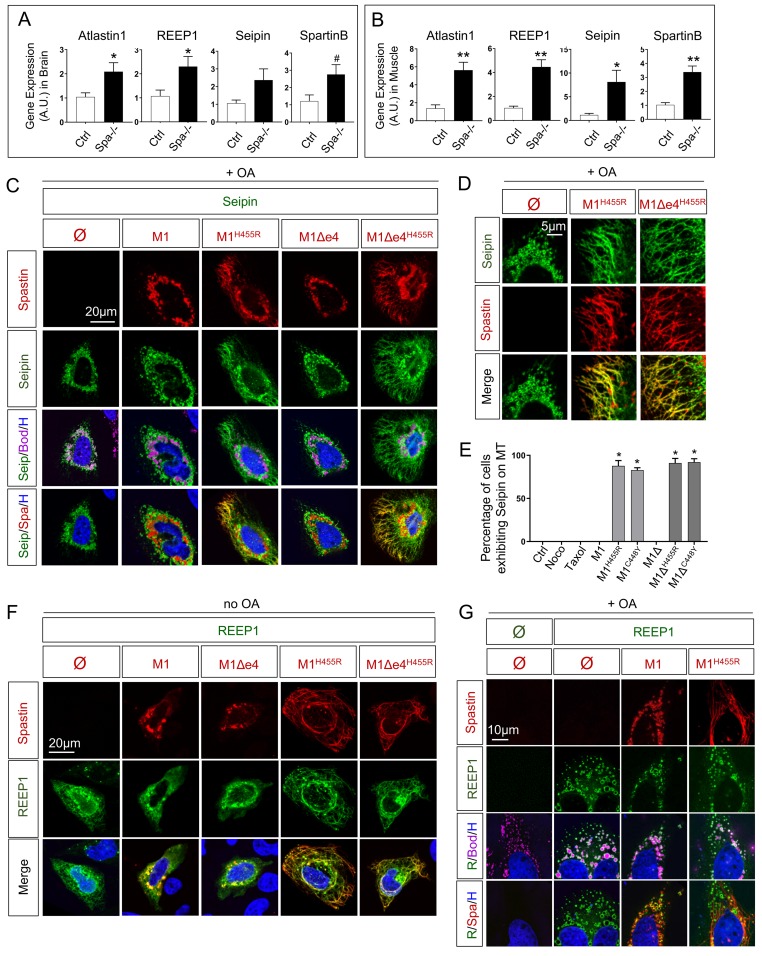
(A) Quantification of REEP1, Atlastin1, Spartin and Seipin transcript by qPCR in 8 months old zebrafish brain. n = 4 animals per group. (B) Quantification of REEP1, Atlastin1, Spartin and Seipin transcript by qPCR in 8 months old zebrafish skeletal muscle. n = 4 animals per group. (C-D) Confocal microscopy images of HeLa cells overexpressing human Seipin with Spastin M1 isoforms (wild-type and mutants) treated with oleic acid (OA) for 18h. Cherry-tagged Spastin appears in red, Seipin in green, LDs (Bodipy) in magenta and nucleus (Hoechst) in blue. (E) Percentage of cells exhibiting tubular ER along MTs. (F-G) Confocal microscopy images of HeLa cells overexpressing human REEP1 with Spastin M1 isoforms in basal conditions (F) or after 18h administration of OA (G). Cherry-tagged Spastin appears in red, REEP1 in green, LDs (Bodipy) in magenta and nucleus (Hoechst) in blue. Bars are mean ± SEM, #