
Figure 6
- ID
- ZDB-IMAGE-200423-90
- Publication
- Murcia-Belmonte et al., 2019 - A Retino-retinal Projection Guided by Unc5c Emerged in Species with Retinal Waves
- All Figures
- Figures for Murcia-Belmonte et al., 2019
|
Figure 6
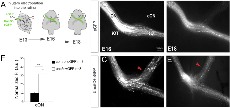
Unc5c Is Sufficient to Guide RGC Axons into the Opposite Optic Nerve
(A) EGFP or Unc5c/GFP-encoding plasmids were electroporated into one eye of E13.5 embryos and axons growing into the opposite optic nerve analyzed at E16.5 or E18.5.
(B–E) E16.5 (B and C) and E18.5 (D and E) embryos electroporated with Unc5c-encoding plasmids (C and E) display an increase in the number of R-R axons (arrowheads) compared to age-matched controls (B and D).
(F) Mean (± SEM) normalized fluorescence intensity in the cON of E16.5 embryos after electroporation of EGFP or Unc5c/GFP-encoding plasmids.
Error bars indicate ± SEM. (∗∗∗p < 0.001, Student’s unpaired t test).
See also