
Figure 3
- ID
- ZDB-IMAGE-200423-86
- Publication
- Murcia-Belmonte et al., 2019 - A Retino-retinal Projection Guided by Unc5c Emerged in Species with Retinal Waves
- All Figures
- Figures for Murcia-Belmonte et al., 2019
|
Figure 3
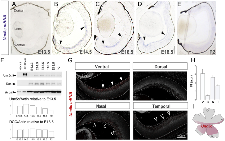
Unc5c Is Expressed in Ventral-Central Retina
(A–E)
(F) Immunoblot detection of Unc5c and Dcc in control (CT) HEK cells and HEK transfected with Unc5c encoding plasmids and in E13.5–P2 retinal lysates. β-actin was used as a loading control. Graphs show levels of Unc5c and Dcc normalized to actin levels. Peak of Unc5c expression is at E14.5.
(G) Representative images of
(H) Mean (± SEM) fluorescent intensity of
(I) Diagram of a whole-mounted retina summarizing the expression of
Error bars indicate ± SEM.
See also