
Image
|
Figure Caption
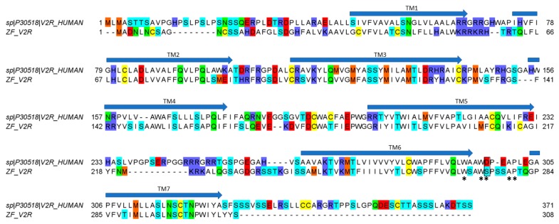
Figure 5
Alignment between hV2R (uniprot entry P30518) and cloned ZF V2R. The substitution of D297 in hV2R with S275 in ZF V2R is bordered in black. Asterisks (*) indicate amino-acid residues that have been suggested to participate and to be important in receptor–ligand interaction. Arrows indicate the seven putative transmembrane domains (TM 1–7).
Acknowledgments
This image is the copyrighted work of the attributed author or publisher, and
ZFIN has permission only to display this image to its users.
Additional permissions should be obtained from the applicable author or publisher of the image.
Full text @ Mar. Drugs