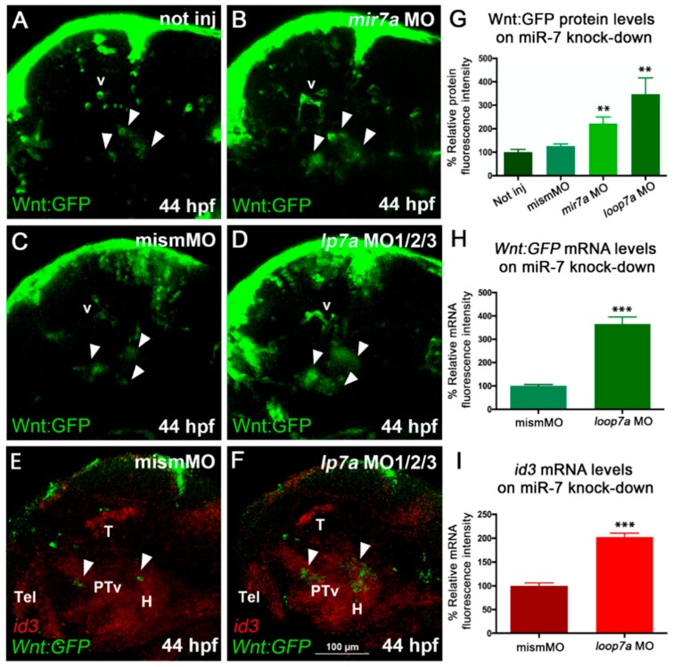
miR-7 knockdown increases Wnt signaling in the zebrafish diencephalon. (A,B,C) Morpholino-mediated knock-down of miR-7 (mature form) increases Wnt-reporter GFP (Green Fluorescent Protein) in the diencephalon (arrowheads) of miR-7 morphants (B) compared with not injected (A) and mismatched MO-injected (C) controls. Increased Wnt-reporter GFP is confirmed in loop7a1/2/3 (immature miR-7) morphants (D). (E,F) Two-color in situ hybridization for id3 (landmark) and Wnt:gfp mRNAs shows that most of the diencephalic Wnt-responsive cells (arrowheads) are located in the ventral posterior tuberculum (PTv) and hypothalamic (H) regions of 44 hpf embryos. Loop7a1/2/3 morphants display increased id3 and gfp expression, compared with controls. All panels show 44 hpf embryonic heads in lateral view, from anterior to the left. Tel: telencephalon; T: tectum; v: vessels. (G,H,I) Quantitative analysis of Wnt reporter in the zebrafish ventral diencephalon. Chart G refers to A–D experimental series; charts H and I refer to E–F experiments. Error bars represent ± SEM. The experiments were repeated thrice. Sample size n = 5 for quantification using Volocity software. Unpaired t-test results are indicated by (**) p < 0.01 and (***) p < 0.001. All panels zoom on the dorsal diencephalic (dd) region of 2 dpf embryos in lateral view, from anterior to the left. Displayed images represent the average phenotype from batches of n = 20 embryos per condition; the experiment was performed in duplicate.
This image is the copyrighted work of the attributed author or publisher, and
ZFIN has permission only to display this image to its users.
Additional permissions should be obtained from the applicable author or publisher of the image.
Full text @ Cells
Your Input Welcome
Thank you for submitting comments. Your input has been emailed to ZFIN curators who may contact you if
additional information is required.
Oops. Something went wrong. Please try again later.