
Fig. 1
- ID
- ZDB-IMAGE-200413-36
- Genes
- Publication
- Villasenor et al., 2019 - Hhex regulates the specification and growth of the hepatopancreatic ductal system
- All Figures
- Figures for Villasenor et al., 2019
|
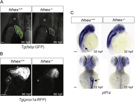
Fig. 1 hhex−/− display impaired liver and pancreas development. (A–C) Analysis of the endodermal phenotypes in hhex−/−. (A) Lateral views of 96 hpf Tg(fabp:GFP) hhex+/+ sibling and hhex−/− larvae. (B) Maximum intensity projections of 96 hpf hhex+/+ sibling and hhex−/− larvae. Ventral views, anterior to the top. hhex−/− display impaired liver development. (C) Dorsal views of whole-mount in situ hybridization for ptf1a expression at 32 and 72 hpf. Compared to stage-matched hhex+/+ siblings, hhex−/− lack exocrine tissue specification, but they display ptf1a neural expression. Black arrows point to pancreatic exocrine tissue. H, heart; L, liver; P, pancreas. Scale bars: 50 μm.
Reprinted from Developmental Biology, 458(2), Villasenor, A., Gauvrit, S., Collins, M.M., Maischein, H.M., Stainier, D.Y.R., Hhex regulates the specification and growth of the hepatopancreatic ductal system, 228-236, Copyright (2019) with permission from Elsevier. Full text @ Dev. Biol.