
Fig 2
- ID
- ZDB-IMAGE-200412-4
- Publication
- Wiles et al., 2020 - Swimming motility of a gut bacterial symbiont promotes resistance to intestinal expulsion and enhances inflammation
- All Figures
- Figures for Wiles et al., 2020
|
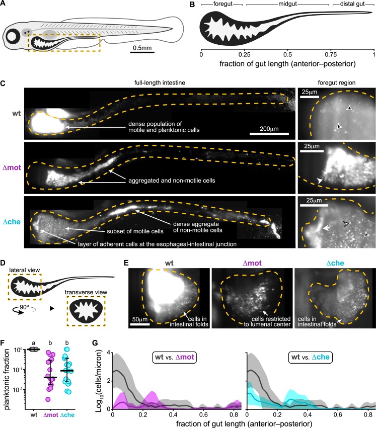
Fig 2
(A) Cartoon of a 6-day-old zebrafish. Dashed box marks intestinal region imaged by LSFM. (B) Anatomical regions of the larval zebrafish intestine. (C) Maximum intensity projections acquired by LSFM showing the spatial organization of wt