IMAGE

FIGURE 10

ID
ZDB-IMAGE-200406-117
Source
Figures for Folgueira et al., 2020
Image
Figure Caption

FIGURE 10

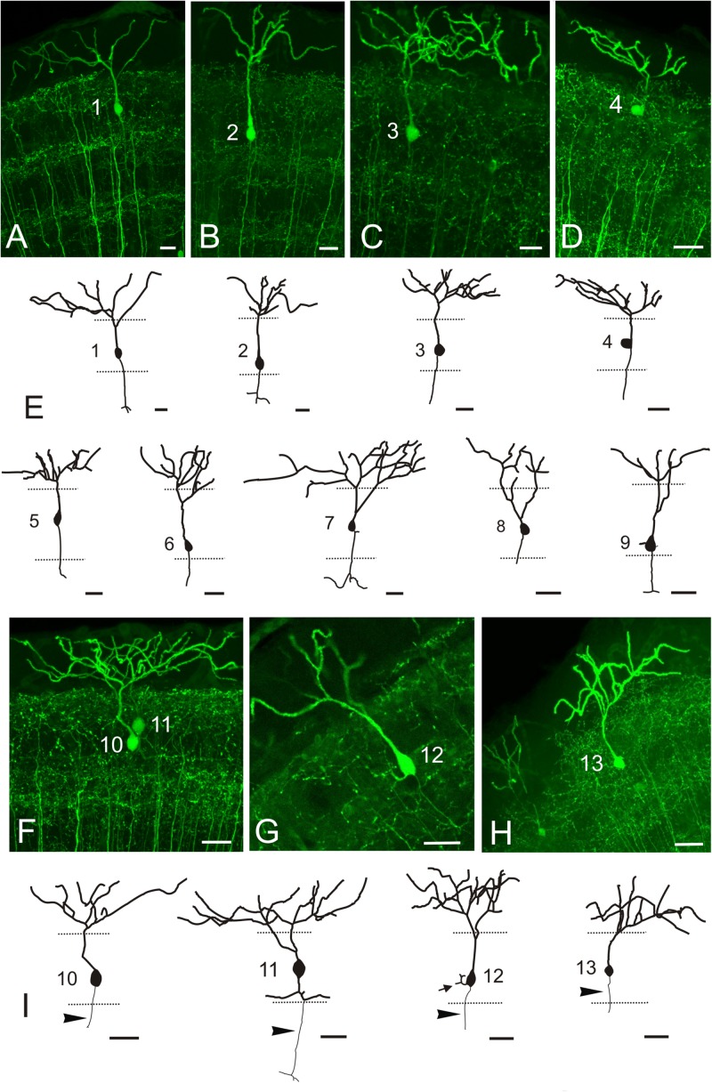
GFP + pyramidal cells in Tg(1.4dlx5a-dlx6a:GFP)ot1. (A–D,F–G) Images showing GFP + pyramidal cells in Tg(1.4dlx5a-dlx6a:GFP)ot1 adult fish. All images are projections from confocal stacks. (E,I) Tracings of 13 pyramidal cells. Apical dendrites are represented with a thicker line than basal ones. Arrowheads in (I) point to axon. All cells are numbered 1-13. Scale bars: 20 μm.

Acknowledgments
This image is the copyrighted work of the attributed author or publisher, and ZFIN has permission only to display this image to its users. Additional permissions should be obtained from the applicable author or publisher of the image. Full text @ Front. Neural Circuits