IMAGE

FIGURE 8

ID
ZDB-IMAGE-200406-115
Source
Figures for Folgueira et al., 2020
Image
Figure Caption

FIGURE 8

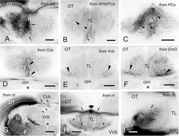
Labeled cells and fibers in TL after DiI application to various regions and nuclei. (A–I), Labeled structures in the TL after unilateral DiI application to the optic tectum (A) pretectal nuclei (B,C), cerebellum (D-F), nucleus isthmi (G,H) and nucleus rostrolateralis (I). (A) Small labeled cells in the ipsilateral TL (arrows) after tracer application to the OT. (B) Very few labeled fibers (arrowheads) in the medial TL after DiI application to the APN and PCe region. (C) Labeled fibers (arrowheads) in the medial TL after tracer application to the paracommissural nucleus. (D–F) Bilaterally labeled fibers (arrowheads) in the ventrolateral TL after unilateral tracer application to the cerebellar corpus (D), cerebellar valvula (E), and granular eminence (F). (G,H) Labeled fibers (arrowheads) after unilateral DiI application to the nucleus isthmi region. Note labeled cells in the optic tectum (white arrowheads) and labeled fibers coursing the intertectal commissure dorsal to TL (thin arrows) in (G) and (H), so as a dorsal longitudinal fascicle (thick arrow) in (H). Note also some fibers in the ipsilateral torovalvular tract (arrowheads), likely labeled by DiI diffusion to nucleus isthmi neighboring areas (see Section “DISCUSSION”), and bilateral labeled fibers in the TL (H). (I), Ipsilateral faintly labeled cells (arrows) in the TL from the rostrolateral nucleus region Asterisk: ventricle. For abbreviations, see the list. Scale bars: Scale bars: 200 μm (G); 100 μm (A-F,H); 20 μm (I).

Acknowledgments
This image is the copyrighted work of the attributed author or publisher, and ZFIN has permission only to display this image to its users. Additional permissions should be obtained from the applicable author or publisher of the image. Full text @ Front. Neural Circuits