
|
Fig. 1
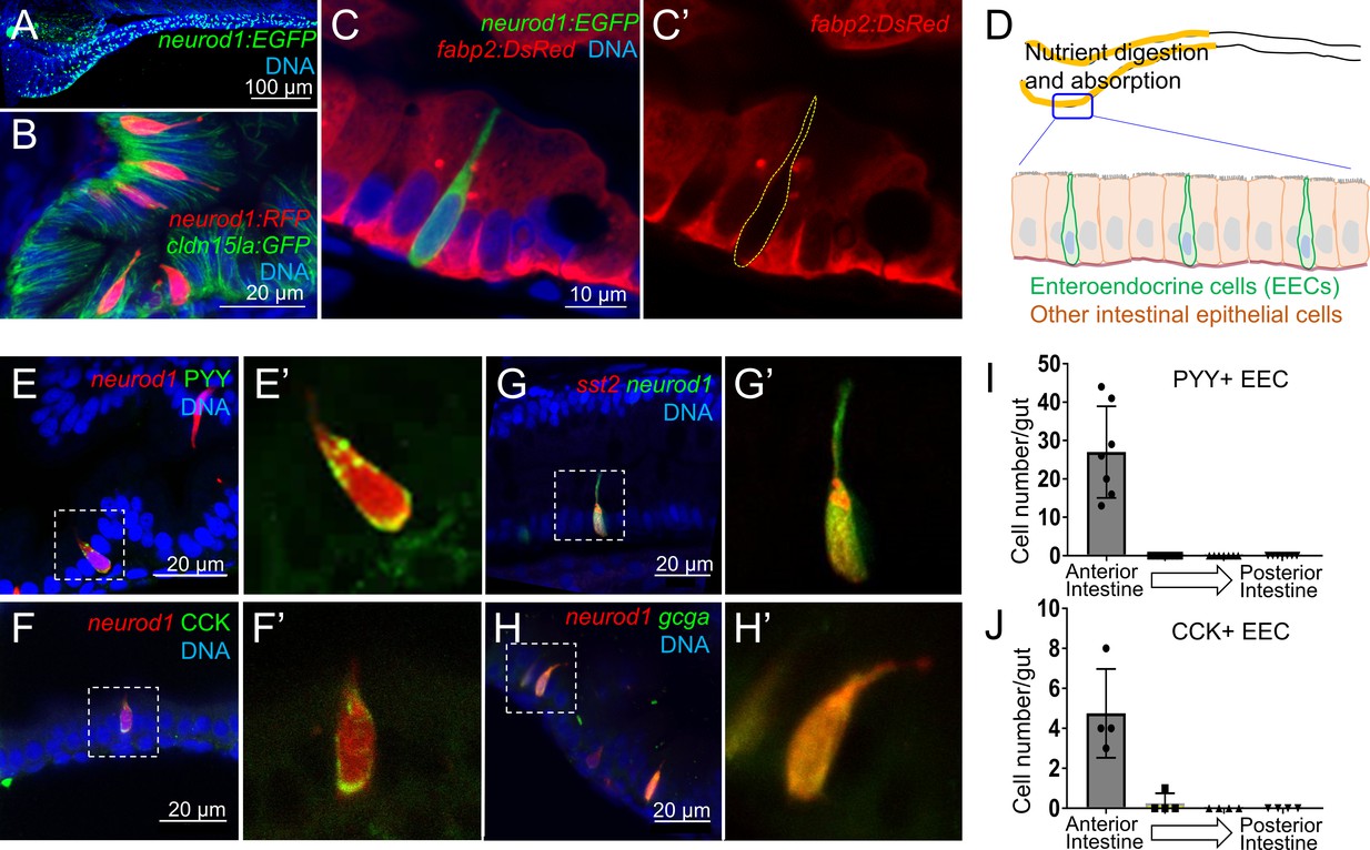
Identification of neurod1+ enteroendocrine cells (EECs) in zebrafish.
( A) Confocal projection of zebrafish EECs marked by the TgBAC(neurod1:EGFP) transgenic line. ( B) Confocal projection of zebrafish EECs marked by Tg(neurod1:RFP). TgBAC(cldn15la:GFP) marks intestinal epithelial cells. ( C) Confocal image of zebrafish EECs marked by TgBAC(neurod1:EGFP) transgenic line. ( C’) Subpanel image of zebrafish enterocyte marked by Tg(fabp2:DsRed). Note that neurod1+ EECs do not express the enterocyte marker fabp2. ( D) Schematic diagram of 6 dpf larval zebrafish intestine. The anterior region of the intestine that is largely responsible for nutrient absorption is highlighted in yellow. ( E–F) Confocal image of neurod1+ EECs stained for PYY (E,) and CCK ( F). ( E’–F’) Zoom view of PYY and CCK positive EECs. ( G–H) Confocal image of neurod1+ EECs expressing somatostatin [marked by Tg(sst2:DsRed) in G] and proglucagon hormones [marked by Tg(gcga:EGFP) in H]. ( G’–H’) Zoom view of sst2 and gcga positive EECs. ( I–J) Quantification of PYY+ (n = 7) and CCK+ (n = 4) EECs in 6 dpf zebrafish intestines.