
Fig. 4
- ID
- ZDB-IMAGE-200323-34
- Genes
- Publication
- Gordon et al., 2019 - Fenestrae-associated protein Plvap regulates the rate of blood-borne proteins passage into the hypophysis
- All Figures
- Figures for Gordon et al., 2019
|
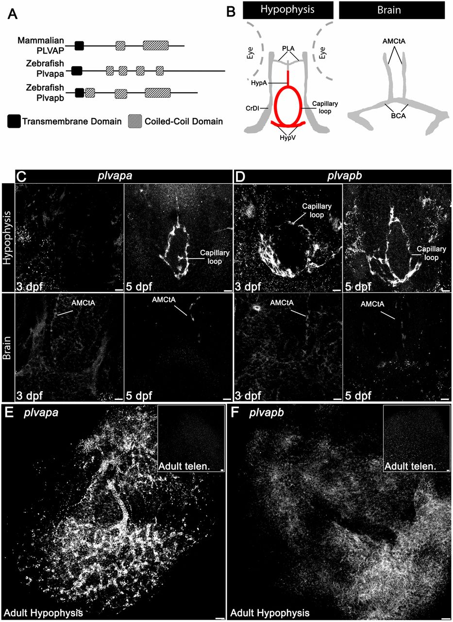
Fig. 4 Developmental expression of Plvap orthologs in the hypophyseal vasculature. (A) Schematic representation of the predicted secondary structure of zebrafish (Plvapa and Plvapb) and mammalian (PLVAP) proteins. (B) A scheme describing the major vasculature of the larval hypophysis and brain. (C,D) Whole-mount fluorescence in situ hybridization of zebrafish larvae at 3 or 5 dpf showing restricted plvapa (C) and plvapb (D) mRNA expression in the hypophyseal but not in the brain vasculature. Scale bars: 5 µm. (E,F) Whole-mount FISH of adult zebrafish showing restricted plvapa (E) and plvapb (F) mRNA expression in the hypophyseal but not in the brain vasculature. Scale bars: 20 µm. AMCtA, anterior (rostral) mesencephalic central artery; BCA, basal communicating artery; CrDI, cranial division of the internal carotid artery; HypA, hypophyseal artery; HypV, hypophyseal vein; PLA, palatocerebral artery. See related Figs S2 and S3.