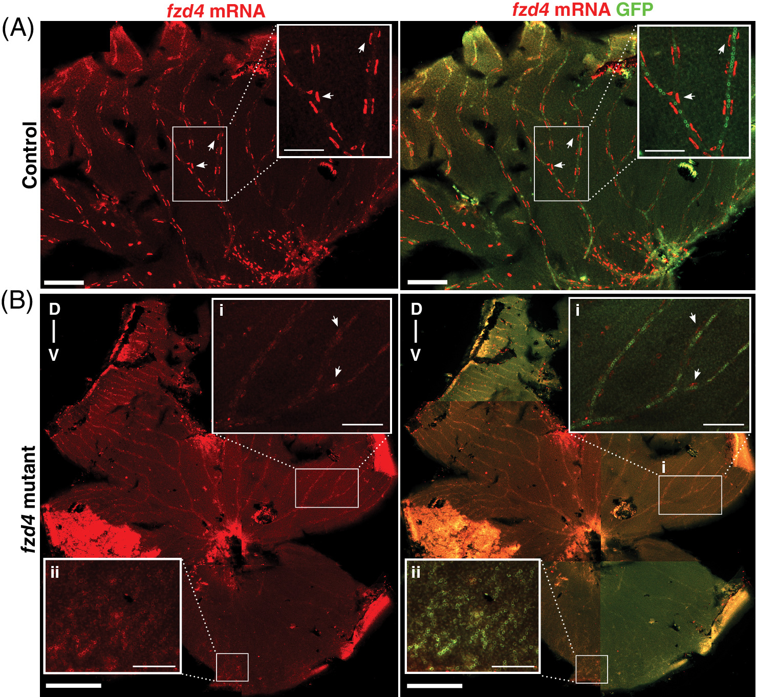
Fig. 7 fzd4 expression is reduced in the adult retinal vasculature of fzd4 mutants. Fluorescence detection of fzd4 mRNA (red) in, A, control and B, fzd4 mutant retinal vasculature. A, In control fish, high fzd4 mRNA levels are detected in discrete regions surrounding the outer border of the blood vessel (inset). Arrowheads point at high levels of fzd4 signal in these discrete regions. Scale bar is 500 μm, inset is 100 μm. B, fzd4 mRNA (red) is reduced in fzd4 mutant fish. This reduction correlates with increased severity of vessel fusion. In the dorsal axis, reduced levels of fzd4 expression are observed (inset B‐i), while no fzd4 expression is detected in the ventral axis (inset B‐ii). Scale bar is 500 μm, inset is 100 μm. Merged panel shows immunofluorescence staining of green fluorescent protein (GFP) (green), which allows for the identification of endothelial cells and the orientation of dissected intraocular sections of the eye. The dorsal/ventral axis is denoted as D‐v
Image
Figure Caption
Figure Data
Acknowledgments
This image is the copyrighted work of the attributed author or publisher, and
ZFIN has permission only to display this image to its users.
Additional permissions should be obtained from the applicable author or publisher of the image.
Full text @ Dev. Dyn.

