IMAGE

FIGURE 3

ID
ZDB-IMAGE-200306-43
Source
Figures for Idilli et al., 2020
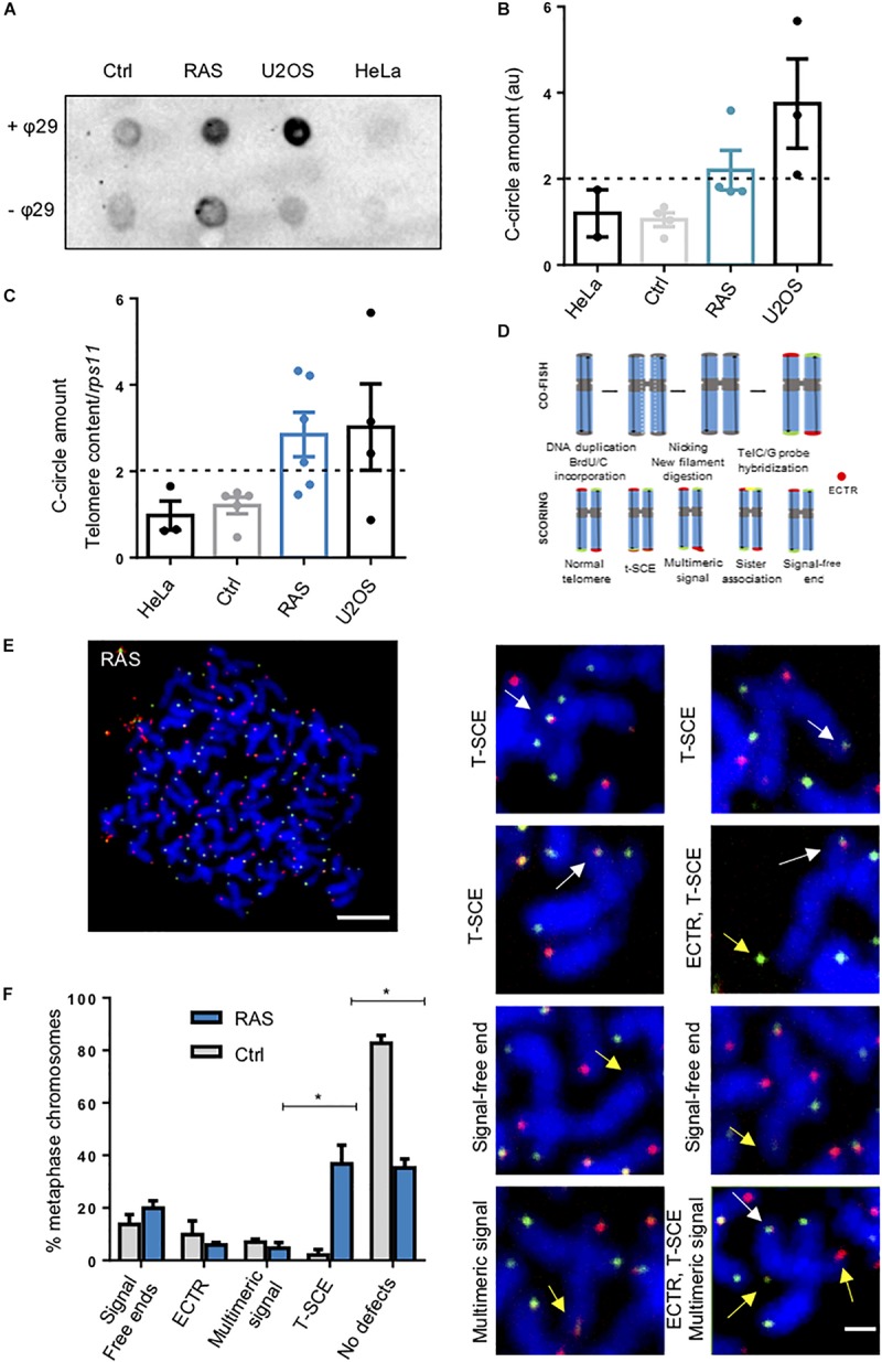
Image
Figure Caption

FIGURE 3

Zebrafish brain tumors are ALT. (A) Representative C-Circle assay by dot blot in one control and one brain tumor compared with telomerase positive HeLa cells and ALT U2OS cells. Reactions without phy29 polymerase (–θ29) were included as a control. (B) Quantitation and analysis of 4 C-Circle assay dot blot. Determination of ALT amount was calculated after subtracting global background and specific –θ29 signal. a.u.: arbitrary unit. (C) C-Circle assay quantified by telomere qPCR. Data are represented as amount of C-circles, normalized to telomere content (TC) and single copy gene (rps11). HeLa and U2OS were added as a reference. Ctrl n = 5; RAS n = 7. The dashed line indicates the level above which ALT activity is considered significant. Whiskers box plots represent median: min to max values. (D) Schematic drawing to describe the procedure for 2-color CO-FISH and the interpretation of telomere status based on the signals. (E) Two-color CO-FISH of a representative metaphase nucleus derived from a RAS brain tumor cell (scale bar 5 μm). The right panels (D) show details of telomeres with T-SCE (white arrows), signal free ends, multimeric signal and/or ECTR (yellow arrows) (scale bar 1 μm). (F) Quantitation of telomeric defects revealed in RAS brain (n = 88 chromosomes) compared with Control brain (n = 56 chromosomes). T-SCE: Telomere Sister Chromatid Exchange; ECTR: Extra-Chromosomal Telomeric Repeat; CO-FISH: Chromosome orientation FISH. Multiple t-test, Holm Sidak methods, *p < 0.05. Bars represent mean ± SEM. See also Supplementary Figure 2.

Figure Data
Acknowledgments
This image is the copyrighted work of the attributed author or publisher, and ZFIN has permission only to display this image to its users. Additional permissions should be obtained from the applicable author or publisher of the image. Full text @ Front Cell Dev Biol