
Fig. S14
- ID
- ZDB-IMAGE-200306-131
- Publication
- Nimura et al., 2019 - Role of Reelin in cell positioning in the cerebellum and the cerebellum-like structure in zebrafish
- All Figures
- Figures for Nimura et al., 2019
|
Fig. S14
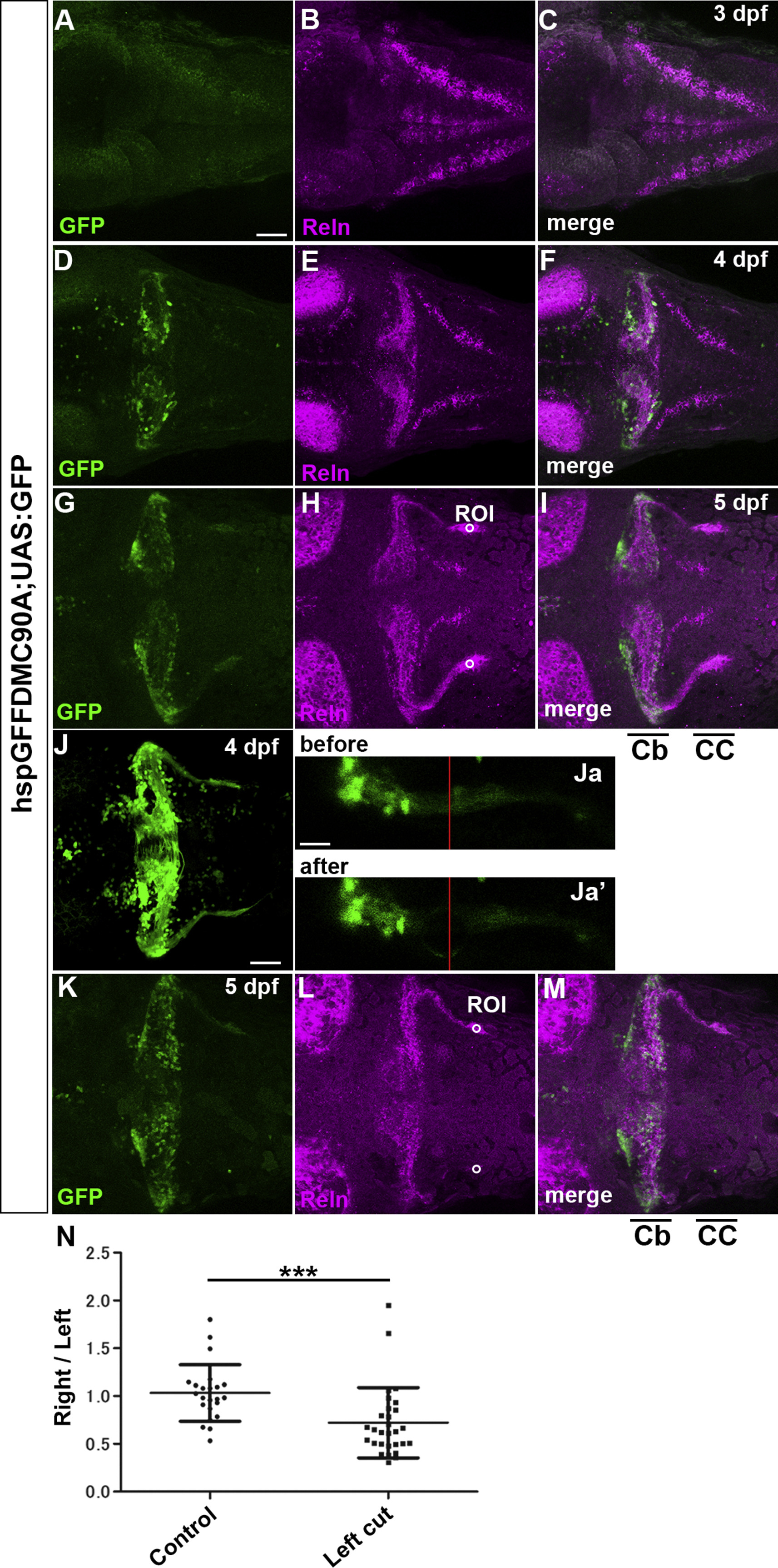
Axon-dependent localization of Reln protein in GCs. (A-I) GC axons and Reln localization. hspGFFDMC90A; UAS:GFP larvae, which express GFP in the soma and axons of GCs, were stained at 3-dpf (A-C), 4-dpf (D-F), and 5-dpf (G-I) with anti-GFP (green) and anti-Reln (magenta) antibodies. Typical data are shown. Reln was detected in or near GC axons in the cerebellum (Cb) and the crista cerebellaris (CC). (J, K) Ablation of GC axons. GC axons in the CC of 4-dpf hspGFFDMC90A; UAS:GFP larvae were ablated by a laser on the left side. (Ja, b) High magnification views of the left GC axons before (Ja) and after the ablation (Ja’). The point of the laser ablation is indicated by red lines. After the ablation, the larvae were reared and subjected to immunostaining with anti-GFP and anti-Reln antibodies at 5 dpf (K). Dorsal views with anterior to the left. Scale bars: 50 μm in A (applies to A-I, K-M); 50 μm in J; 20 μm (applies to Ja, Jb). (N) Fluorescence intensity of Reln signals on the right and left side (ROI, region of interest) in control (H, n= 23) and laser-treated larvae (L, n = 29) was measured. The ratio of the signal on the right to left side in each larva was calculated and plotted in a graph. The ratio was significantly different in the laser-treated larvae (***p< 0.001; Mann-Whitney test).
Reprinted from Developmental Biology, 455(2), Nimura, T., Itoh, T., Hagio, H., Hayashi, T., Di Donato, V., Takeuchi, M., Itoh, T., Inoguchi, F., Sato, Y., Yamamoto, N., Katsuyama, Y., Del Bene, F., Shimizu, T., Hibi, M., Role of Reelin in cell positioning in the cerebellum and the cerebellum-like structure in zebrafish, 393-408, Copyright (2019) with permission from Elsevier. Full text @ Dev. Biol.