
IMAGE
Fig. S12
- ID
- ZDB-IMAGE-200306-129
- Publication
- Nimura et al., 2019 - Role of Reelin in cell positioning in the cerebellum and the cerebellum-like structure in zebrafish
- All Figures
- Figures for Nimura et al., 2019
Image
|
Figure Caption
Fig. S12
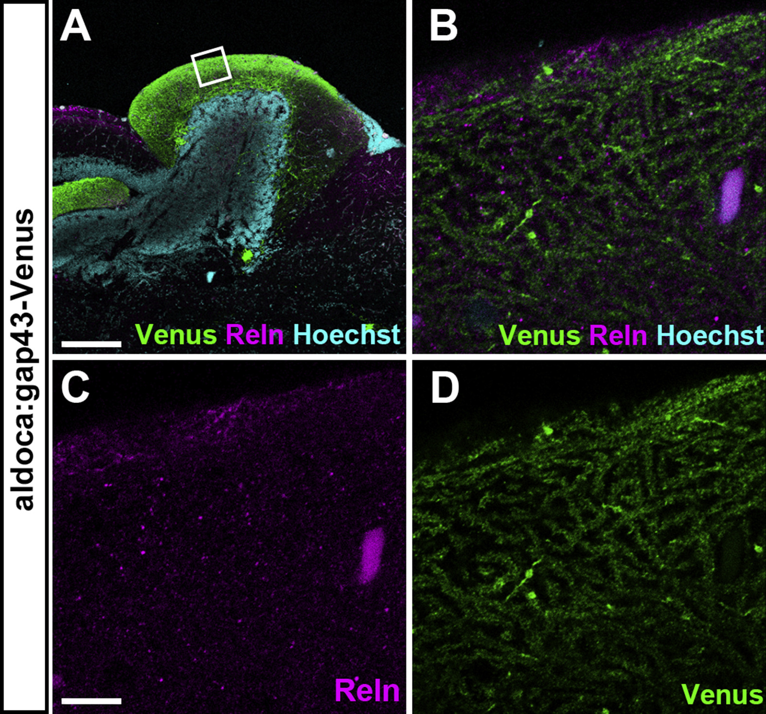
Reln is not colocalized with dendrites of PCs in the ML of the cerebellum. Sagittal sections of the brain from adult Tg(aldoca:gap43-Venus) fish, which express Venus in PCs, were stained with anti-GFP (Venus) and anti-Reln (magenta) antibodies, and Hoechst (cyan). Confocal optical sections (0.93 μm thickness). (B-D) High magnification images of the box in A. Note that Reln is not colocalized with PC dendrites. Scale bars: 200 μm in A; 10 μm in C (applies to B-D).
Acknowledgments
This image is the copyrighted work of the attributed author or publisher, and
ZFIN has permission only to display this image to its users.
Additional permissions should be obtained from the applicable author or publisher of the image.
Reprinted from Developmental Biology, 455(2), Nimura, T., Itoh, T., Hagio, H., Hayashi, T., Di Donato, V., Takeuchi, M., Itoh, T., Inoguchi, F., Sato, Y., Yamamoto, N., Katsuyama, Y., Del Bene, F., Shimizu, T., Hibi, M., Role of Reelin in cell positioning in the cerebellum and the cerebellum-like structure in zebrafish, 393-408, Copyright (2019) with permission from Elsevier. Full text @ Dev. Biol.