
Fig. S10
- ID
- ZDB-IMAGE-200306-127
- Publication
- Nimura et al., 2019 - Role of Reelin in cell positioning in the cerebellum and the cerebellum-like structure in zebrafish
- All Figures
- Figures for Nimura et al., 2019
|
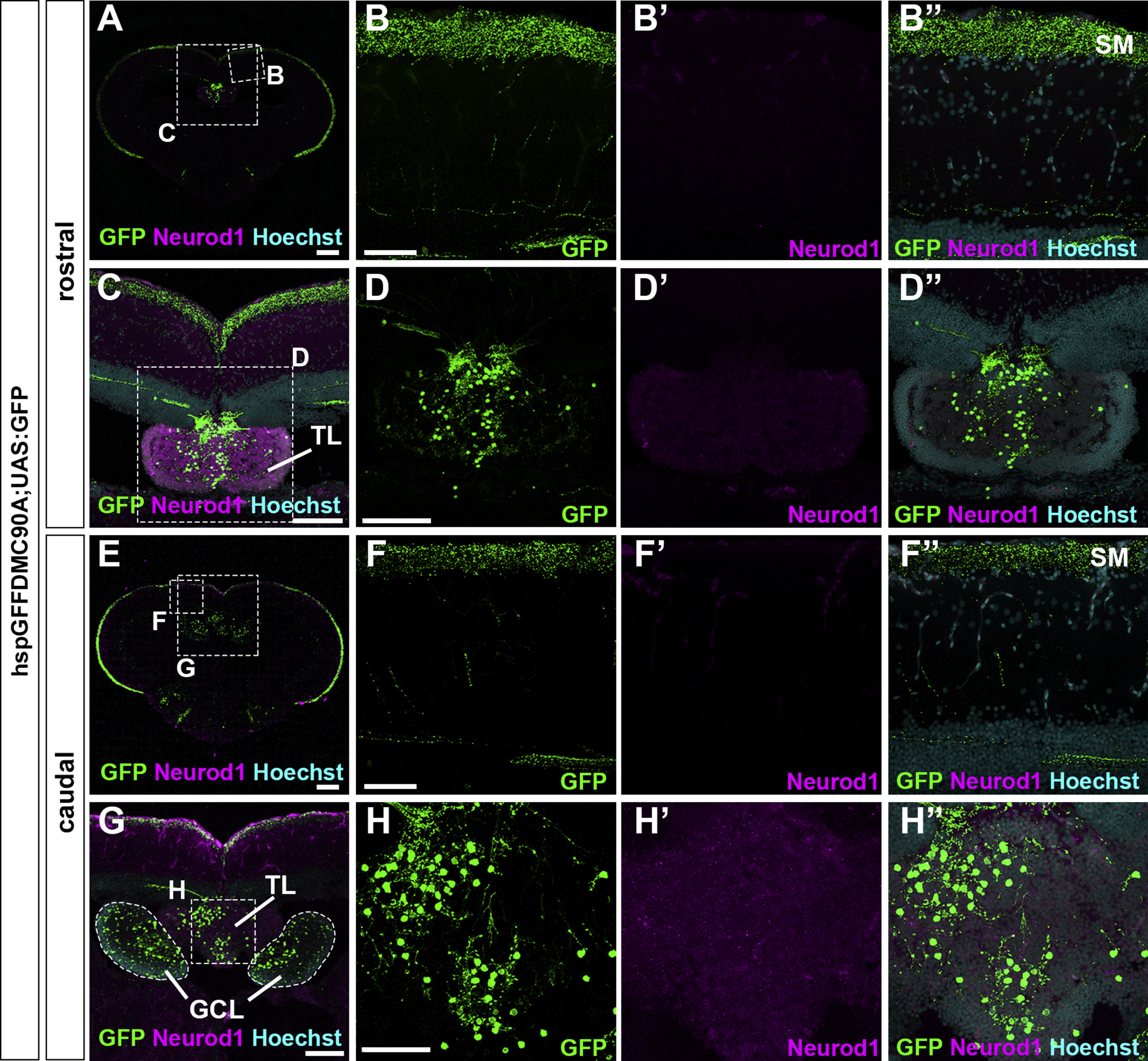
Fig. S10
Granule cells in the TL send their axons to the SM in the tectum. Cross sections of a tectum region of the brain from adult hspGFFDMC90A; UAS:GFP fish, which express GFP specifically in GCs in the TL and the cerebellum, were stained with anti-GFP (green) and anti-Neurod1 (magenta) antibodies, and Hoechst (cyan). (B, C, D, F, G, H) High magnification images of the boxes in A, C, E, and G. Note that GFP was detected in the SM, the TL, and the GCL. The abbreviations are described in the legend of Fig. 1. Scale bars: 200 μm in A and E, 100 μm in C, D (applies to D′, D″), and G, 50 μm in B (applies to B, B′, B″); F (applies to F, F′, F″); and H (applies to H, H′, H″).
Reprinted from Developmental Biology, 455(2), Nimura, T., Itoh, T., Hagio, H., Hayashi, T., Di Donato, V., Takeuchi, M., Itoh, T., Inoguchi, F., Sato, Y., Yamamoto, N., Katsuyama, Y., Del Bene, F., Shimizu, T., Hibi, M., Role of Reelin in cell positioning in the cerebellum and the cerebellum-like structure in zebrafish, 393-408, Copyright (2019) with permission from Elsevier. Full text @ Dev. Biol.