
Fig. S4
- ID
- ZDB-IMAGE-200306-121
- Publication
- Nimura et al., 2019 - Role of Reelin in cell positioning in the cerebellum and the cerebellum-like structure in zebrafish
- All Figures
- Figures for Nimura et al., 2019
|
Fig. S4
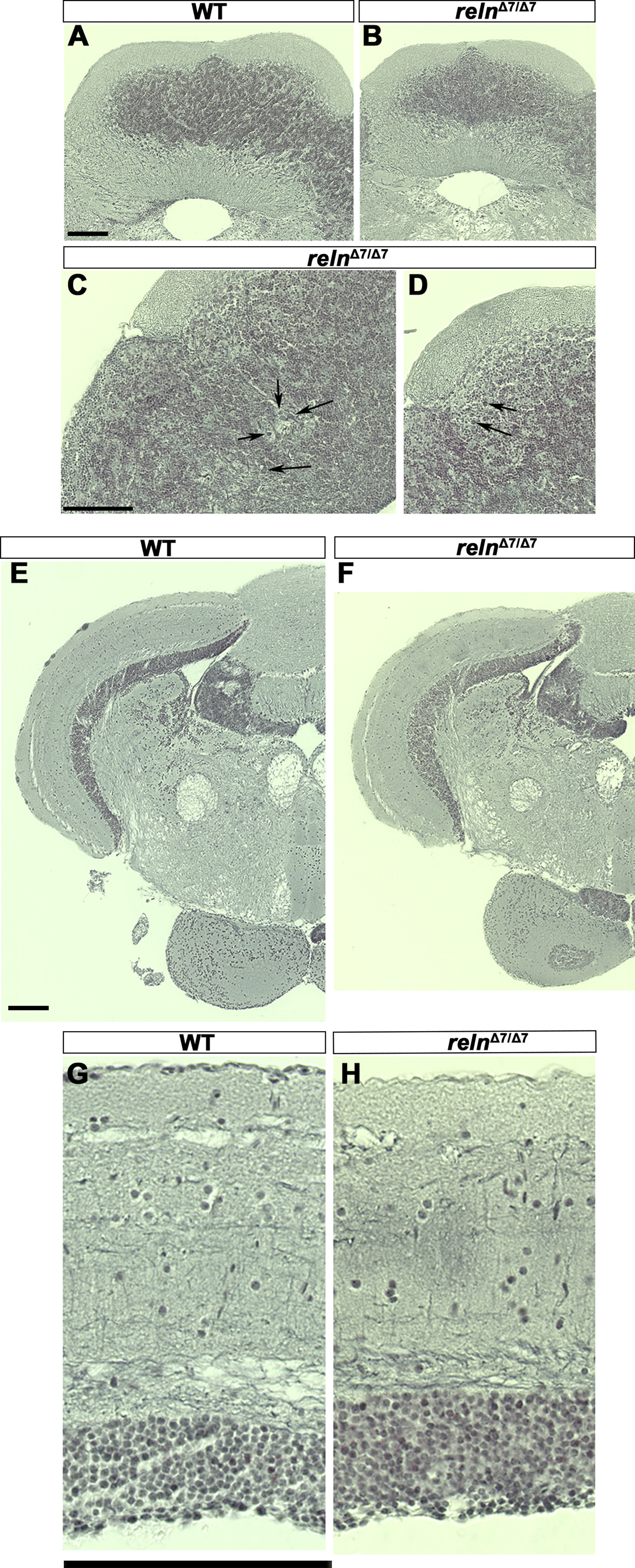
Morphology of the cerebellum and the mesencephalic tectum in reln mutants. Cross sections of the cerebellum (A-D) and the mesencephalon (E-H) of adult WT (A, E, G, n= 2) and relnΔ7/Δ7 mutant (B, C, D, F, and H, n= 2) fish were stained by the Bodian silver impregnation method, which visualizes neuronal fibers (gray) and cell nuclei (purple). (C, D) High magnification images of the relnΔ7/Δ7 mutant cerebellum. Arrows indicate ectopic neurons that have a large soma and are probably PCs in the GCL. (G, H) High magnification images of the WT and relnΔ7/Δ7 mutant tectum. Note that no gross abnormalities in the tectum of the reln mutant were observed. Scale bars: 100 μm in A (applies to A, B); C (applies to C, D); E (applies to E, F); and G (applies to G, H).
Reprinted from Developmental Biology, 455(2), Nimura, T., Itoh, T., Hagio, H., Hayashi, T., Di Donato, V., Takeuchi, M., Itoh, T., Inoguchi, F., Sato, Y., Yamamoto, N., Katsuyama, Y., Del Bene, F., Shimizu, T., Hibi, M., Role of Reelin in cell positioning in the cerebellum and the cerebellum-like structure in zebrafish, 393-408, Copyright (2019) with permission from Elsevier. Full text @ Dev. Biol.