
Fig. 3
- ID
- ZDB-IMAGE-200306-113
- Genes
- Antibodies
- Publication
- Nimura et al., 2019 - Role of Reelin in cell positioning in the cerebellum and the cerebellum-like structure in zebrafish
- All Figures
- Figures for Nimura et al., 2019
|
Fig. 3
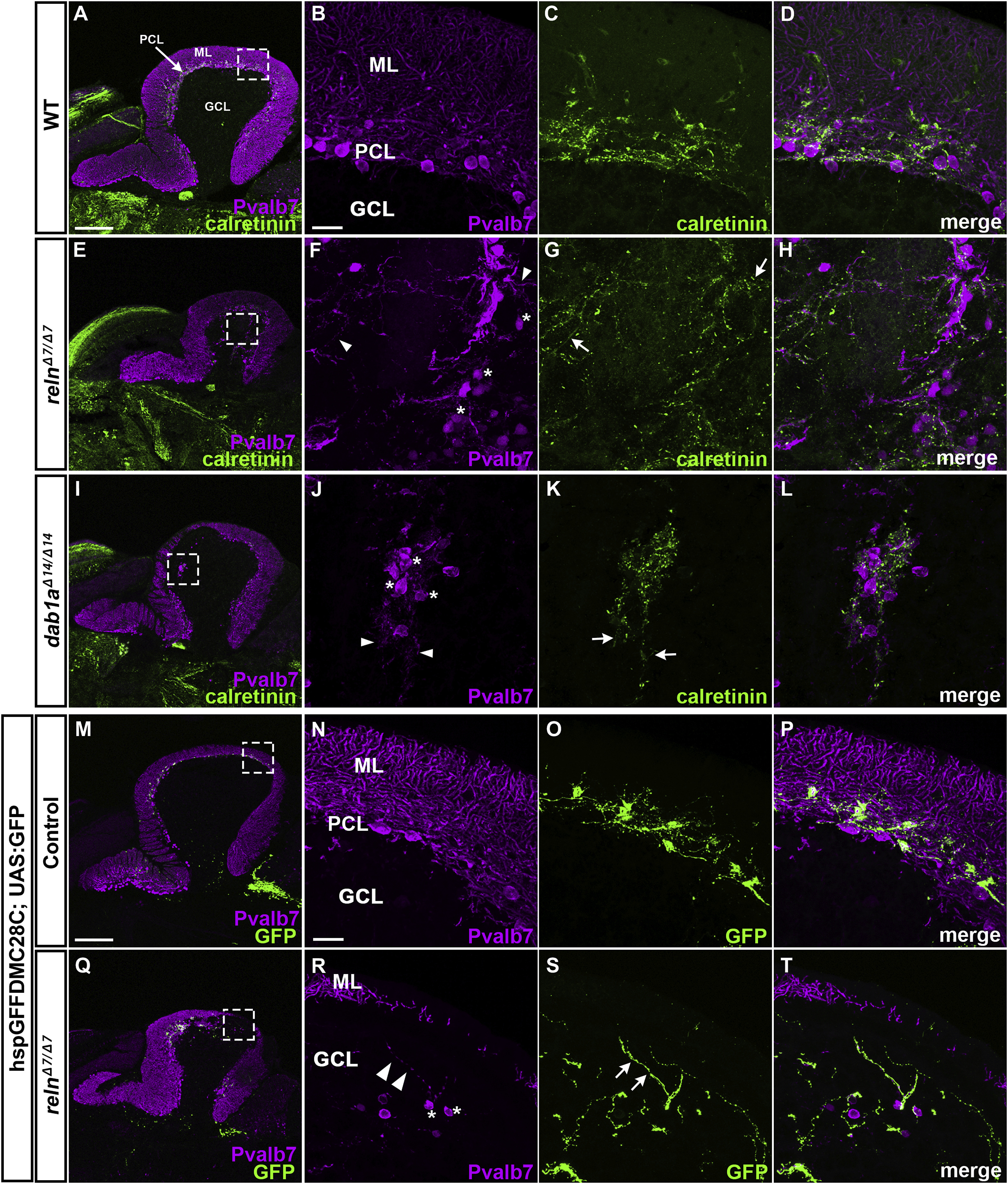
Projection of climbing fibers (CFs) to ectopic PCs in reln and dab1amutants. (A–L) Calretinin-immunoreactive (CR-ir+) axonal projections to PCs. Sagittal sections of adult (92-dpf) WT (A-D, n= 4) and relnΔ7/Δ7mutant (E-L, n= 4) brains were stained with anti-Pvalb7 (magenta) and anti-calretinin antibodies (green). (B-D, F–H, J-L) High magnification images of the boxes in A, E, and I. Typical images are shown. CR-ir+ axons projected to ectopic PCs located in the GCL in reln mutant cerebella, but not in WT cerebella. (M–T) CF projections to ectopic PCs. Sagittal section of adult (96 dpf) hspGFFDMC28C (28C); UAS:GFP fish brains, which express GFP in the CFs (axons of the neurons in the inferior olivary nuclei), harboring WT (control, n= 4) or homozygous reln mutant (relnΔ7/Δ7, n= 4) alleles were stained with anti-Pvalb7 (magenta), and anti-GFP (green) antibodies. GFP+ CFs projected to ectopic PCs located in the GCL in reln mutant cerebella, but not in WT cerebella. Ectopic somata and dendrites of PCs are indicated by asterisks and arrowheads, respectively. CR-ir+ and 28C; UAS:GFP+ axons projecting to the ectopic Purkinje cells are indicated by arrows. The abbreviations are described in the legend for Fig. 1. Scale bars: 200 μm in A (applies to A, E, and I); 20 μm in B (applies to B-D, F–H, J-L); 200 μm in M (applies to M and Q); 20 μm in N (applies to N-P and R-T).
Reprinted from Developmental Biology, 455(2), Nimura, T., Itoh, T., Hagio, H., Hayashi, T., Di Donato, V., Takeuchi, M., Itoh, T., Inoguchi, F., Sato, Y., Yamamoto, N., Katsuyama, Y., Del Bene, F., Shimizu, T., Hibi, M., Role of Reelin in cell positioning in the cerebellum and the cerebellum-like structure in zebrafish, 393-408, Copyright (2019) with permission from Elsevier. Full text @ Dev. Biol.