
|
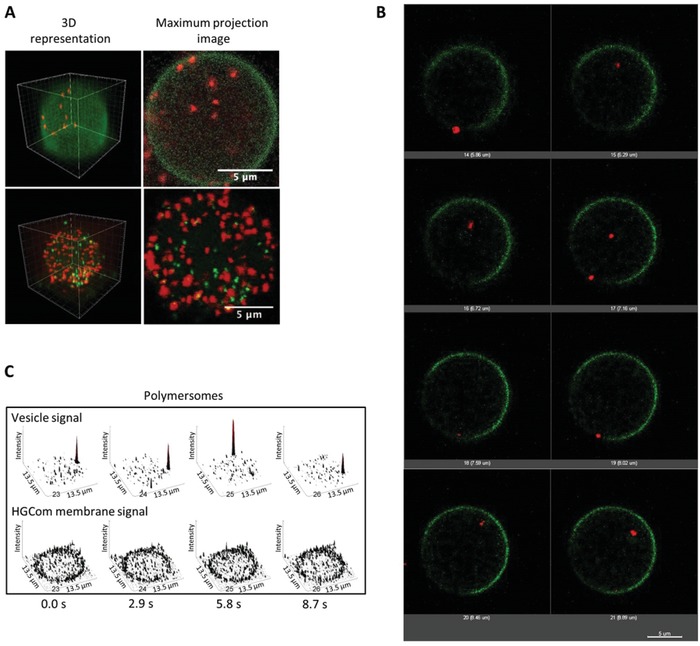
Figure 4
Subcompartmentalization within E‐GPMVs. A) 3D reconstructions (left) and maximum intensity projection images of Z stacks (right) of E‐GPMVs with multicompartment architecture. Top: SRB‐loaded polymersomes inside E‐GPMVs equipped with Lck‐GFP. Red: signal of SRB‐loaded polymersomes. Green: signal of Lck‐GFP membrane protein. Bottom: SRB‐loaded polymersomes and CF‐loaded polymersomes inside E‐GPMVs. Red: signal of SRB encapsulated inside polymersomes. Green: signal of CF encapsulated inside polymersomes. B) Single plane projections of different Z positions during E‐GPMV Z‐stacking process by CLSM. Z interval = 1 µm. Red: SRB loaded polymersomes. Green: Lck‐GFP membrane protein. C) Single plane recordings of PMOXA6‐PDMS44‐PMOXA6 polymersomes inside E‐GPMVs measured by CLSM. See Movies S3–S8 in the Supporting Information.