
|
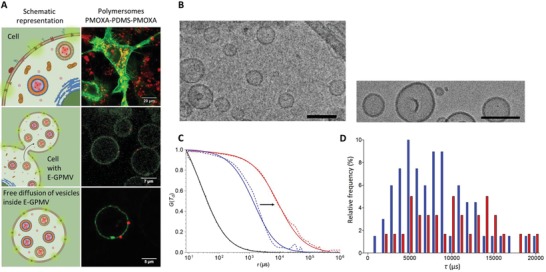
Figure 3
Transfer of cytosolic components from the cellular cytosol into E‐GPMVs. A) Left column: Top) Schematic representation of HepG2 cells with enhanced membrane and cytosolic content. Central) Illustration of the formation of several equipped‐GPMV with multicompartment structure (E‐GPMV) from the donor cells. Bottom) One isolated E‐GPMV with microcompartment structure. Right column: Top) CLSM micrograph of HepG2 cells containing membrane protein LcK‐GFP (Green) and SRB‐loaded PMOXA6‐PDMS44‐PMOXA6 polymersomes (Red), Scale bar 20 µm. Central) CLSM micrograph of several E‐GPMV, Scale bar 7 µm. Bottom) CLSM micrograph of one isolated E‐GPMV simultaneously containing Lck‐GFP protein and SRB‐loaded PMOXA6‐PDMS44‐PMOXA6 polymersomes, Scale bar 5 µm. See Movies S3–S8 in the Supporting Information. CLSM Controls are available in the Supporting Information. B) Cryogenic transmission electron micrograph of PMOXA6‐PDMS44‐PMOXA6 polymersomes in PBS pH 7.4. Scale bar represents 200 nm (left) and 50 nm (right). C) FCS analysis of SRB‐loaded PMOXA6‐PDMS44‐PMOXA6 polymersomes in PBS pH 7.4 (Blue) and SRB‐loaded PMOXA6‐PDMS44‐PMOXA6 polymersomes within E‐GPMVs (Red), Free SRB at pH 7.4 PBS (Black). Arrowhead indicates the change in diffusion time from free SRB polymersomes once they are encapsulated in E‐GPMVs. Dotted line—experimental auto correlation curves, full line—fit. Curves normalized to 1 to facilitate comparison. D) Frequency distribution of diffusion times taken from individual FCS measurements: SRB‐loaded PMOXA6‐PDMS44‐PMOXA6 polymersomes in PBS pH 7.4 (Blue) and SRB‐loaded PMOXA6‐PDMS44‐PMOXA6 polymersomes within E‐GPMVs (Red) (