
Figure 1
- ID
- ZDB-IMAGE-200227-8
- Publication
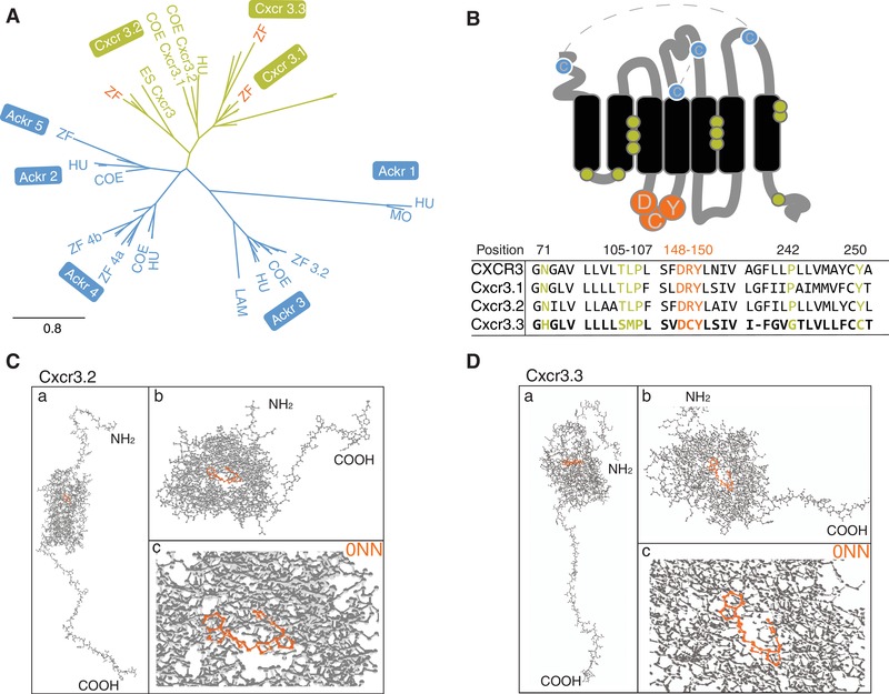
- Sommer et al., 2019 - Frontline Science: Antagonism between regular and atypical Cxcr3 receptors regulates macrophage migration during infection and injury in zebrafish
- All Figures
- Figures for Sommer et al., 2019
|
Figure 1