IMAGE

Figure 5

ID
ZDB-IMAGE-200213-30
Source
Figures for Gregson et al., 2019
Image
Figure Caption

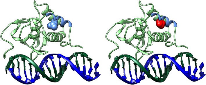
Figure 5

(A) Wild‐type (WT) SMAD9 MH1 domain (light green ribbon with helix‐1 in light blue) binding the DNA helix (dark blue/dark green). L22 is shown in blue space filling. (B) L22P, shown in red space filling, is predicted to severely disrupt the structure of the MH1 domain. Supplemental Movie S2 in File S1: 3‐dimensional rotating image.

Acknowledgments
This image is the copyrighted work of the attributed author or publisher, and ZFIN has permission only to display this image to its users. Additional permissions should be obtained from the applicable author or publisher of the image. Full text @ J. Bone Miner. Res.