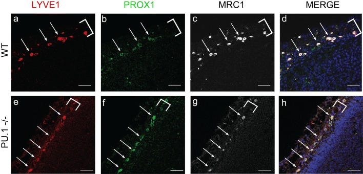
IMAGE

Fig. 4

ID
ZDB-IMAGE-200213-14
Source
Figures for Shibata-Germanos et al., 2019
Image
Figure Caption

Fig. 4

Mouse LLECs develop independent of the transcription factor PU.1. ad As detected by IHC, E15.5 wild-type mice have cells that co-express LYVE1 (a, red) PROX1 (b, green) and MRC1 (c, white) in the developing meninges (white bracket). DAPI labels nuclei blue in the merged image (d). White arrows indicate cells with these three markers. Scale = 50 µm. n = 2 brains. e–h) The meninges (white bracket) of E15.5 PU.1 knockout siblings contain many cells (white arrows) that co-express LYVE1 (e, red), PROX1 (f, green), and MRC1 (g, white). Scale = 50 µm. n = 3 brains

Acknowledgments
This image is the copyrighted work of the attributed author or publisher, and ZFIN has permission only to display this image to its users. Additional permissions should be obtained from the applicable author or publisher of the image. Full text @ Acta Neuropathol.