
|
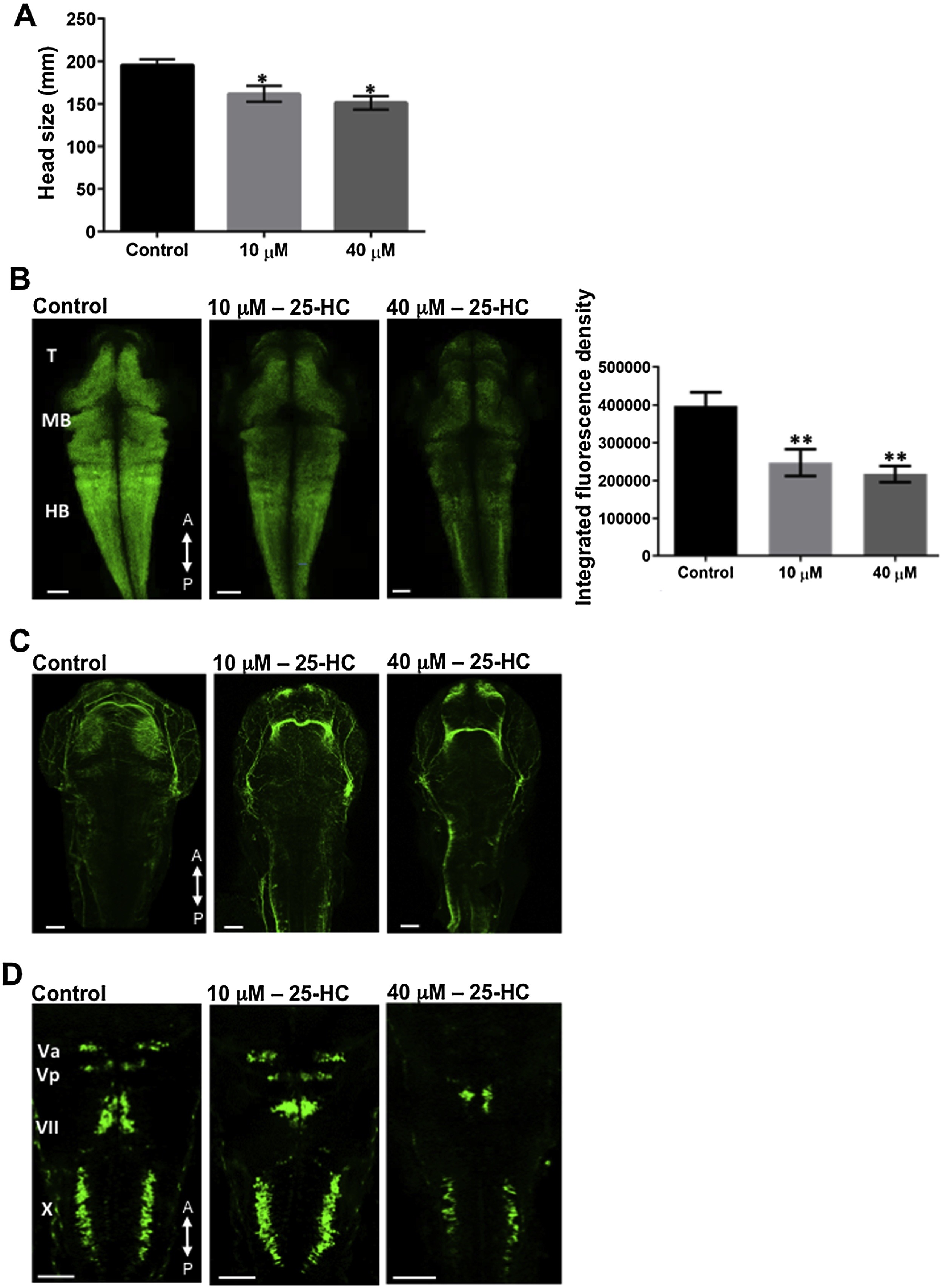
Fig. 4
25-HC administration alters brain development in zebrafish: (A) Head size comparison between the treated and un-treated larvae at 2 dpf. Characterizing the larval brains at 2 dpf using Tg(huc:GFP) (B) and Tg(Isl1:GFP) (D) lines showed a decrease in the neuronal population and a defective development of the cranial nerves, in a dose dependent manner. (C) Larvae stained for acetylated α-tubulin at 2 dpf, showed reduced axon network clusters in the brain. All comparisons are performed with control, each trial has a n of 7 for control and treated over 4 trials, * represents a significance of p < 0.05, ** represents a significance of p < 0.01. Significance was determined using one-way ANOVA and all data are represented as Mean ± SEM. T: Telencephalon, MB: Midbrain, HB, Hindbrain. A, P represent anterior and posterior side of the head respectively.