
Fig. 8
- ID
- ZDB-IMAGE-200203-10
- Genes
- Publication
- Zhu et al., 2019 - Zebrafish prmt5 arginine methyltransferase is essential for germ cell development
- All Figures
- Figures for Zhu et al., 2019
|
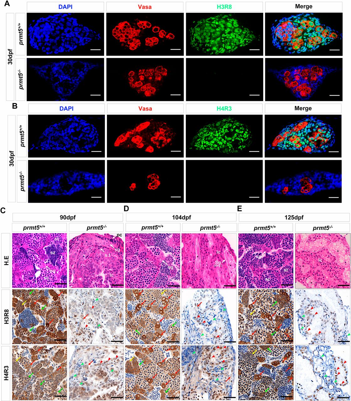
Fig. 8
The deletion of prmt5 in zebrafish results in reduction of H3R8me2s and H4R3me2s. (A,B) Vasa (red), H3R8me2s (green) (A), H4R3me2s (green) (B) and DNA (DAPI staining; blue) in prmt5+/+ testes (n=6) and prmt5−/− gonads (n=6) at 1 mpf. No H3R8me2s-positive cells (A) or no H4R3me2s-positive cells (B) were detected in prmt5−/− gonads. (C-E) Hematoxylin and Eosin staining, and H3R8me2s and H4R3me2s staining in wild-type sibling controls (prmt5+/+) and prmt5 homozygous mutants (prmt5−/−). At 90 dpf (C), the testes of prmt5+/+ zebrafish showed typical testicular morphology, with abundant spermatogonia (sg), spermatocytes (sc), spermatid (st) and spermatozoa (sz). s.t, seminiferous tube. The gonads of prmt5−/− had pyknotic cells (pc), but few spermatogonia (sg) and no spermatocytes (sc). At 104 dpf (D), in the gonads of prmt5−/− zebrafish, lumens (indicated with white dashed lines) began to enlarge, while remaining germ cells were replaced by Leydig cells (lc) and Sertoli cells (SC). The spermatogonia in the lumens gradually degenerated (Sg deg., asterisk). At 125 dpf (E), in the gonads of prmt5−/− zebrafish, lumens (indicated with white dashed lines) underwent further enlargement, germ cells were replaced by Leydig cells (lc) and the spermatogonia in the lumens degenerated. Spermatogonia, red arrows; spermatocytes, green arrows; Leydig cells, green arrowheads; Sertoli cells, red arrowheads. Scale bars: 20 μm.