Image
Figure Caption

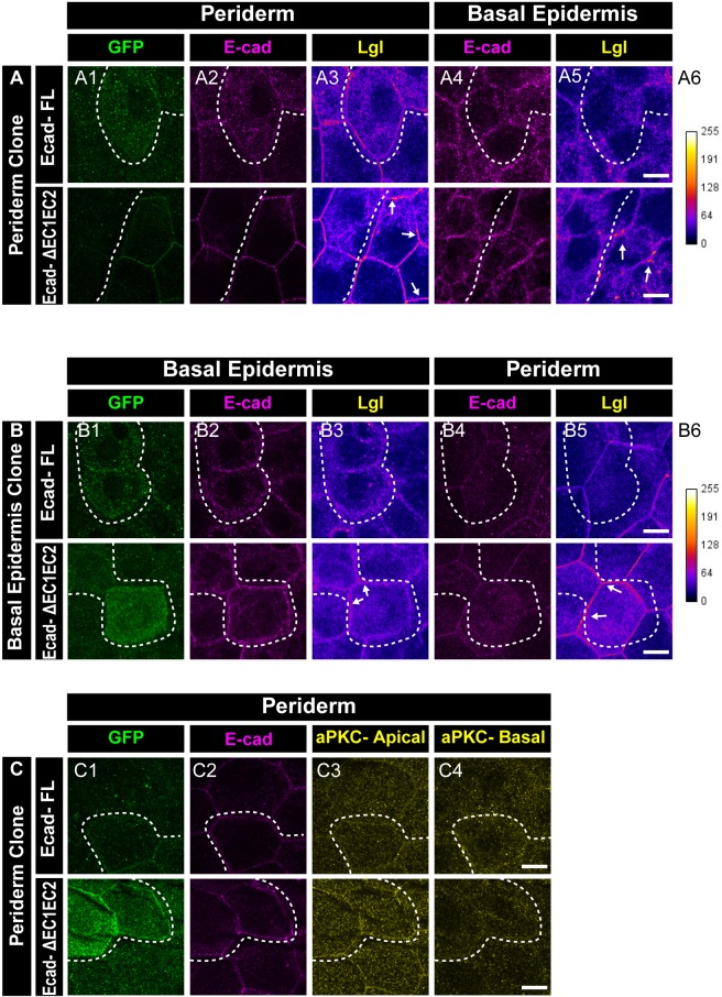
Figure 5—figure supplement 4. EC1-EC2 domain of E-cadherin is required to maintain Lgl2 levels in the epidermis.

Confocal images showing expression and localisation of GFP tagged E-cadherin-FL and ΔEC1-EC2-Ecad (green) (A1, B1, C1) and localisation of E-cadherin (magenta) (A2, A4, B2, B4, C2), Lgl2 (A3, A5, B3, B5) and aPKC (C3, C4). Lgl staining in the periderm (A3, B5) or basal epidermis (B3, A5) is shown as heatmap with calibration bar provided for reference (A6, B6). Arrow shows high levels of Lgl in the ΔEC1-EC2-Ecad expressing cells or juxtaposed cells (A3, A5, B3, and B5). Dotted lines indicate the position of the clone in the periderm (A1–A3 and C1-C4) or basal epidermis (B1–B3) and corresponding region in basal epidermis (A4–A5) or periderm (B4–B5), respectively. Scale bar represents 10 µm in (A5, B5, C4).

Acknowledgments
This image is the copyrighted work of the attributed author or publisher, and ZFIN has permission only to display this image to its users. Additional permissions should be obtained from the applicable author or publisher of the image. Full text @ Elife