Image
Figure Caption

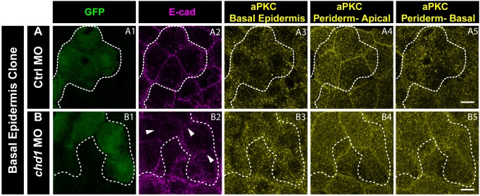
Figure 5—figure supplement 2. <italic>e-cadherin</italic> knockdown in the basal epidermis does not have any effect on aPKC localisation in the periderm or basal epidermis.

Immunostaining of embryos having Ctrl MO (A) and cdh1 MO (B) clones marked by GFP (A1, B1) and showing e-cadherin knockdown (A2, B2), aPKC localisation in the basal epidermis (A3, B3) and the apical domain (A4, B4) as well as basal domain of the peridermal cells (A5, B5) lying above the basal epidermal clone. Note that there is no effect on aPKC localisation in the periderm when e-cadherin is knocked down in the basal epidermis. Dotted lines mark the basal epidermal clone and the peridermal region above the clone. Arrowheads (in B2) mark the loss of E-cadherin in the morphant clone. Scale bar equals to 10 µm (A5, B5).

Acknowledgments
This image is the copyrighted work of the attributed author or publisher, and ZFIN has permission only to display this image to its users. Additional permissions should be obtained from the applicable author or publisher of the image. Full text @ Elife