
IMAGE
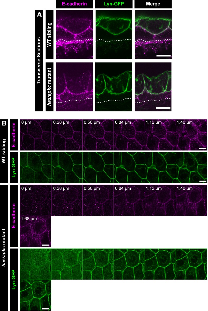
Figure 2—figure supplement 2.
- ID
- ZDB-IMAGE-200125-28
- Publication
- Arora et al., 2020 - Stepwise polarisation of developing bilayered epidermis is mediated by aPKC and E-cadherin in zebrafish
- All Figures
- Figures for Arora et al., 2020
Image
|
Figure Caption
Figure 2—figure supplement 2.
Transverse section through the dorsal head epidermis at 48hpf showing localisation of E-cadherin (magenta) and Lyn-GFP (green) along the apicobasal axis in WT siblings and
Acknowledgments
This image is the copyrighted work of the attributed author or publisher, and
ZFIN has permission only to display this image to its users.
Additional permissions should be obtained from the applicable author or publisher of the image.
Full text @ Elife