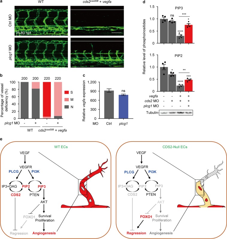
PIP3 reduction is mainly caused by PLCγ mediated PIP2 hydrolysis. Representative confocal images (a) and quantitative analysis (b) of trunk vessel phenotypes of WT or cds2 mutant embryos with vegfa OE and with or w/o plcg1 MO. The ISV number counted from 20–22 embryos per group is shown on the top (b). c Relative vegfa mRNA level of cds2 mutants with vegfa OE injected with ctrl or plcg1 MO. vegfa expression level was determined at 2 h post heatshock induction and normalized to WT embryos without vegfa OE. n = 3 samples, 15–20 embryos pooled for each sample. dplcg1 knockdown partially restored PIP2 and PIP3 level in cds2-deficient endothelium with vegfa OE. Western blotting analysis on a-Tubulin serves as the internal control for cell amounts in each sample collected during lipid quantitation analysis. n = 4 samples per group. e Working model of VEGFA-triggered vessel regression on CDS2-deficient endothelium. The outcome of VEGFA signaling can be reversed from angiogenesis to vessel regression, which is dependent on CDS2-controlled PIP2 and PIP3 availability and FOXO1 signaling activation. Scale bar, 100 μm. Error bars, mean ± SEM. *P < 0.05; **P < 0.01; ***P < 0.001; ****P < 0.0001; ns, not significant (P ≥ 0.05). See also Supplementary information, Fig. S8
This image is the copyrighted work of the attributed author or publisher, and
ZFIN has permission only to display this image to its users.
Additional permissions should be obtained from the applicable author or publisher of the image.
Full text @ Cell Res.
Your Input Welcome
Thank you for submitting comments. Your input has been emailed to ZFIN curators who may contact you if
additional information is required.
Oops. Something went wrong. Please try again later.