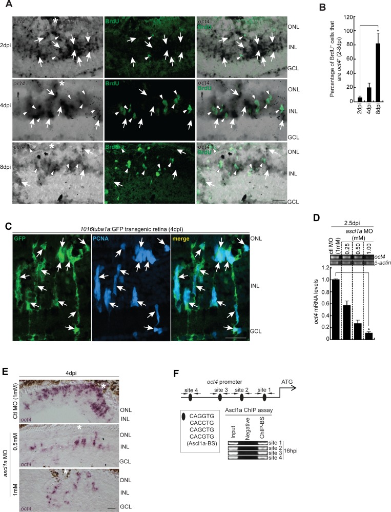
Increased co-localization of oct4 with MGPCs towards later stages of retina regeneration, and the effect of ascl1a knockdown on oct4 expression.
(A, B) BF and IF confocal microscopy images of retinal cross sections show increased oct4 mRNA in BrdU+ MGPCs towards the later stages of retina regeneration (A), which is quantified (B); *P < 0.0001 (t test), N = 4. (A) White arrowheads mark BrdU+ cells and white arrows mark oct4+ cells in (A). (C) IF confocal microscopy images of retinal cross sections from 1016tuba1a:GFP transgenic retina show the GFP expression in PCNA+ MGPCs at 4 dpi. White arrows mark co-labeled PCNA and GFP expression. (D) RT-PCR (upper) and qPCR (lower) analysis of oct4 mRNA in ascl1a knockdown retina at 2.5 dpi. (E) BF microscopy images of retinal cross sections show the decline in oct4 mRNA with increasing concentrations of ascl1a MO at 4 dpi. (F) The oct4 promoter schematic reveals the typical Ascl1a-BSs (upper) and the retinal ChIP assays confirm the physical binding of Ascl1a at the typical BSs (lower) in 16 hpi retina. Ctl MO is control MO. Error bars are SD. (A, C, E) Scale bars, 10 μm; the asterisk marks the injury site, GCL, ganglion cell layer; INL, inner nuclear layer; ONL, outer nuclear layer (A, C, E).
This image is the copyrighted work of the attributed author or publisher, and
ZFIN has permission only to display this image to its users.
Additional permissions should be obtained from the applicable author or publisher of the image.
Full text @ Life Sci Alliance
Your Input Welcome
Thank you for submitting comments. Your input has been emailed to ZFIN curators who may contact you if
additional information is required.
Oops. Something went wrong. Please try again later.