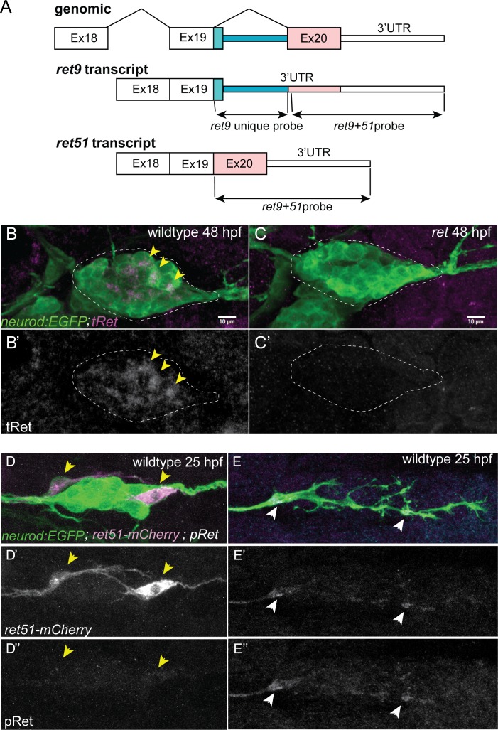
(A) Schematic of ret genomic locus, spliced ret9 and ret51 isoforms, and anti-sense mRNA probe targets for fluorescence in situ hybridization in Figure 1. ret9 in situ probe contains the unique portion of exon 19, specific to this isoform, and intron 19–20 sequence, whereas ret9+51 in situ probe encompasess exon 20 (contained within ret9 transcript 3’UTR) and 3’UTR common to both isoforms. (B,C) Whole-mount immunolabeling for total Ret protein of wild-type sibling (B) and rethu2846 mutant (C) with anti-tRet antibody at 48 hpf. neurod:EGFP transgene marks all neurons in the pLL ganglion (outlined). tRet immunoreactivity (arrowheads) is present a subset of wild-type neurons but is completely absent from the rethu2846 mutant. N = 8 wild-type sibs and N = 6 mutants from two independent experiments. (D,E) Whole-mount immunolabeling of wild-type siblings with anti-phospho-Ret (p905) antibody. neurod:egfp-positive embryos were injected with 5 pg of neurod5kb:RET51-mCherry fusion plasmid and fixed at 25 hpf. Anti-pRet antibody recognizes overexpressed RET51-mCherry fusion protein. Panels show immunolabeling of pLL ganglion (D) and pioneer growth cones (E) in wild-type embryo with pRet antibody. Note absence of the signal in the ganglion or cell bodies of neurons that express Ret51-mCherry (yellow arrowheads). However, pRet signal was reliably detected in growth cones (white arrowheads). N = 12 cells from two independent experiments.
Acknowledgments
This image is the copyrighted work of the attributed author or publisher, and
ZFIN has permission only to display this image to its users.
Additional permissions should be obtained from the applicable author or publisher of the image.
Full text @ Elife
Your Input Welcome
Thank you for submitting comments. Your input has been emailed to ZFIN curators who may contact you if
additional information is required.
Oops. Something went wrong. Please try again later.