
Image
|
Figure Caption
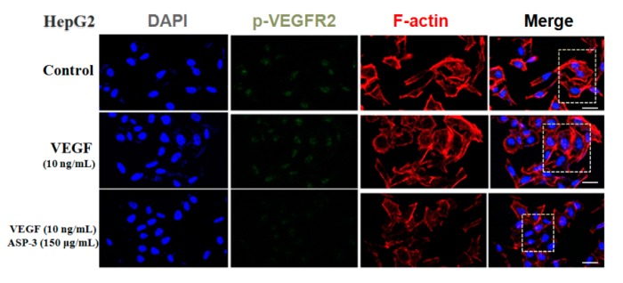
Figure 7
ASP-3 reduced VEGFR2 phosphorylation in HepG2 cells. Immunofluorescence staining was used to evaluate the distribution of VEGFR2 phosphorylation. They were stained with DAPI (blue), fluorescent secondary antibody of phospho-VEGFR2 VEGFR2 (green), and phalloidin (red), respectively. The immunofluorescence profile was visualized under a confocal fluoresce (scale bar: 20 µm).
Acknowledgments
This image is the copyrighted work of the attributed author or publisher, and
ZFIN has permission only to display this image to its users.
Additional permissions should be obtained from the applicable author or publisher of the image.
Full text @ Mar. Drugs