IMAGE

Figure 10

ID
ZDB-IMAGE-191230-1566
Source
Figures for Zhang et al., 2019
Image
Figure Caption

Figure 10

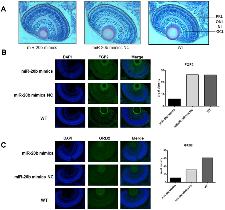
The zebrafish model confirms the inhibitory effect of miR-20b. (A) Observation of retinal tissue sections at 96hpf. We found that in the miRNA-20b mimic group, there were fewer cells in the photoreceptor layer than in the control and wild groups, and the arrangement of cells was disordered. PRL: Photoreceptor layer, ONL: Outer nuclear layer, INL: Inner nuclear layer, GCL: Ganglion cell layer. (B, C) Immunofluorescence detected decreased FGF2 and GRB2 brightness compared to controls. Relative fluorescence values were analyzed using Imagej software (*P<0.05).

Acknowledgments
This image is the copyrighted work of the attributed author or publisher, and ZFIN has permission only to display this image to its users. Additional permissions should be obtained from the applicable author or publisher of the image. Full text @ Aging (Albany NY)