IMAGE

Figure 3

ID
ZDB-IMAGE-191230-154
Source
Figures for Jung et al., 2019
Image
Figure Caption

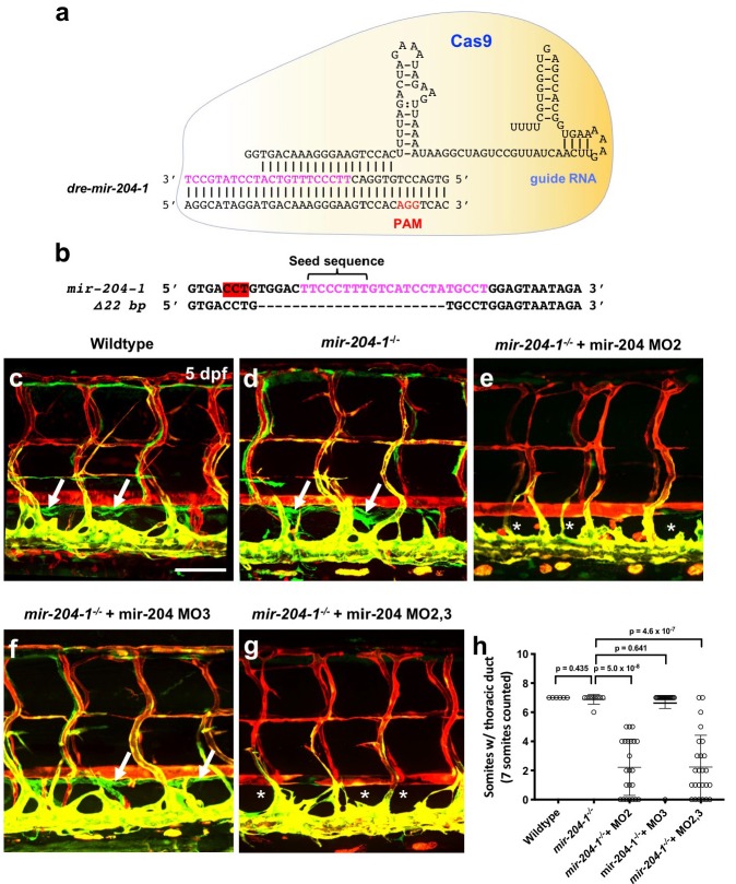
Figure 3 Lymphatic differentiation defects in mir-204-deficient animals.

(a) Quantitative TaqMan RT-PCR measurement of the relative levels of mature mir-204 in one dpf mir-204–1-/- mutants and wildtype siblings, normalized to wild type levels. (b,c) Confocal images of 2 dpf Tg(mrc1a:eGFP)y251, Tg(kdrl:mCherry)y171 mir-204–1-/- mutant (c) or MO2-injected mir-204–1-/- mutant (d) animals. (d) Quantitation of the number of PL sprouts in animals as in panels b and c. Scale bar: 100 μm (b). All graphs are analyzed by t-test and the mean ± SD is shown.

Figure Data
Acknowledgments
This image is the copyrighted work of the attributed author or publisher, and ZFIN has permission only to display this image to its users. Additional permissions should be obtained from the applicable author or publisher of the image. Full text @ Elife