IMAGE

Fig. 5

ID
ZDB-IMAGE-191230-1536
Source
Figures for Xiao et al., 2019
Image
Figure Caption

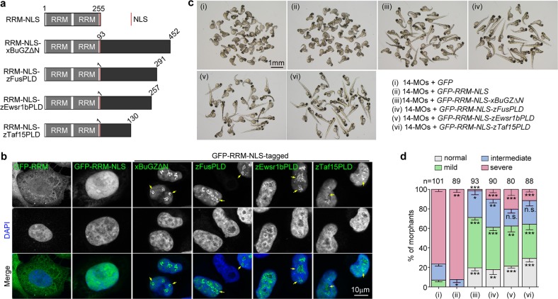
Fig. 5 The IDR of zRbm14b can be functionally replaced with other phase separation domains in vivo.

a Diagrams of chimeric proteins examined. RRM, the RRM region of zRbm14b; NLS, the nuclear localization signal of large T antigen; xBuGZΔN, the IDR of xBuGZ; zFusPLD, zEwsr1bPLD, or zTaf15PLD, the PLD domain of zebrafish Fus, Ewsr1b, or Taf15 (also see Supplementary Fig. 4e). Numbers indicate amino acid positions in each intact protein. b Subcellular localization of the indicated GFP-tagged proteins. HeLa cells were transfected with the intact plasmids used for in vitro mRNA productions (c) for 48 h and fixed with 4% paraformaldehyde. Nuclear DNA was stained with DAPI. Arrows indicate representative nuclear foci. c, d The phase separation domains of xBuGZ, zFus, zEwsr1b, and zTaf15 were partially redundant to the IDR of zRbm14b. Zebrafish embryos at the one-cell stage were co-injected with 14-MOs (8 ng per embryo) and the indicated in vitro-transcribed mRNA (300 pg mRNA per embryo) (also see Supplementary Fig. 4d, f) and imaged at 72 hpf (c). The quantification results (d), based on the criteria and examples in Fig. 1e and represented as mean ± SD, were from three independent experiments. Student’s t-test against the GFP mRNA-injected populations: n.s., no significance; *P < 0.05; **P < 0.01; ***P < 0.001. Total numbers of embryos analyzed are listed over the histogram

Acknowledgments
This image is the copyrighted work of the attributed author or publisher, and ZFIN has permission only to display this image to its users. Additional permissions should be obtained from the applicable author or publisher of the image. Full text @ Cell Discov