
Fig. 4
- ID
- ZDB-IMAGE-191104-19
- Genes
- Antibodies
- Publication
- Liu et al., 2019 - Chemokine signaling links cell-cycle progression and cilia formation for left-right symmetry breaking
- All Figures
- Figures for Liu et al., 2019
|
Fig. 4
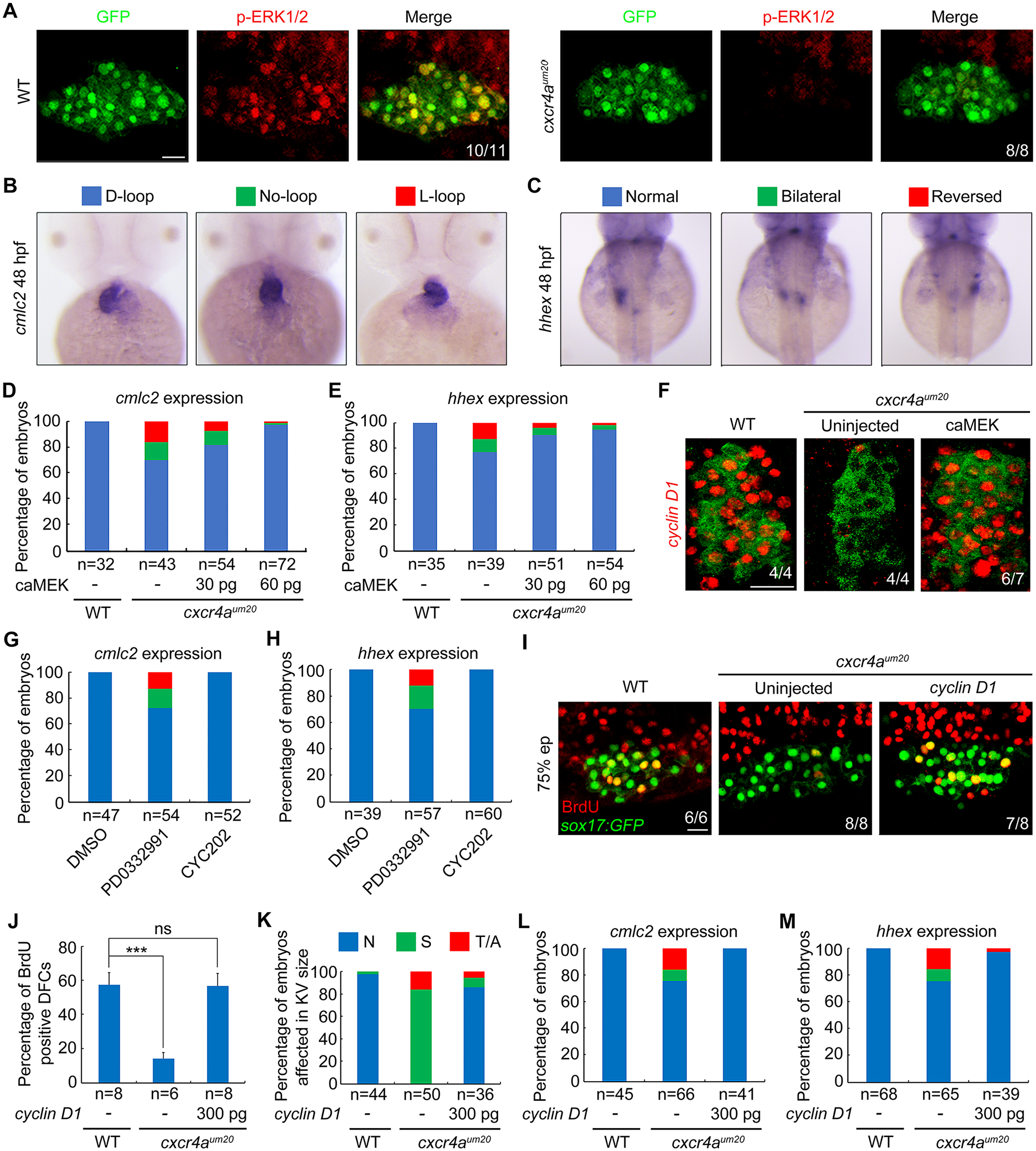
Cxcr4 promotes Cyclin D1 expression through ERK signaling during DFC proliferation.
(A) ERK1/2 phosphorylation levels were dramatically decreased in cxcr4aum20 mutants. WT and cxcr4a-deficient Tg(sox17:GFP) embryos were harvested at the 75% epiboly stage and subjected to immunostaining for p-ERK1/2 (red) and GFP (green). All embryos are shown in dorsal views with anterior to the top. Scale bar, 20 μm. (B–E) caMEK mRNA overexpression in DFCs rescued L–R patterning defects in cxcr4aum20 mutants. Different types of heart looping and liver laterality at 48 hpf in cxcr4aum20 mutants following midblastula injection of different caMEK mRNA doses were visualized by cmlc2 and hhex expression (B and C). The ratios are shown in (D) and (E). Underlying data can be found in S1 Data. (F) Cxcr4a-deficient Tg(sox17:GFP) embryos were injected with 60 pg caMEK mRNA at the 256-cell stage and then harvested at the 75% epiboly stage for fluorescence in situ hybridization experiments with cyclin D1 (red) and GFP (green) probes. Dorsal views with anterior to the left. Scale bar, 20 μm. (G–H) WT embryos were treated with 0.5 μM PD0332991 or 0.2 μM CY202 from the shield stage to bud stage and then analyzed for L–R patterning defects at 48 hpf by in situ hybridizations with cmlc2 and hhex probes. The proportion of treated embryos exhibiting each type of heart looping and liver laterality is shown in (G) and (H). Underlying data can be found in S1 Data. (I–J) Reintroduction of cyclin D1 into DFCs relieves DFC proliferation defects in cxcr4aum20 mutants. Cxcr4a-deficient Tg(sox17:GFP) embryos were injected with or without 300 pg cyclin D1 mRNA at the 256-cell stage, followed by coimmunostaining with anti-BrdU (red) and anti-GFP (green) antibodies at the 75% epiboly stage. Representative images are shown in (I), and the percentage of BrdU-positive DFCs is indicated in (J). Scale bar, 20 μm. Student t test, ***P < 0.001. Underlying data can be found in S1 Data. (K–M) The reduced ratios of embryos affected in KV size (K) and cmlc2 (L) or hhex (M) expression show that DFC-specific overexpression of cyclin D1 rescued the defects of KV formation (K) and L–R patterning (L and M) in cxcr4a mutants. Underlying data can be found in S1 Data. BrdU, bromodeoxyuridine; caMEK, constitutively activated version of MEK; cmlc2, cardiac myosin light chain 2; DFC, dorsal forerunner cell; ERK, extracellular regulated MAP kinase; GFP, green fluorescent protein; hhex, hematopoietically expressed homeobox; hpf, hours postfertilization; KV, Kupffer’s vesicle; L–R, left–right; ns, no significant difference; p-ERK1/2, phosphorylated ERK1/2; sox, SRY-box transcription factor; Tg, transgene; WT, wild-type